The discovery, explained · Chemist no. 188 of 350
Osamu Shimomura and GFP: The Jellyfish That Lit Up Biology
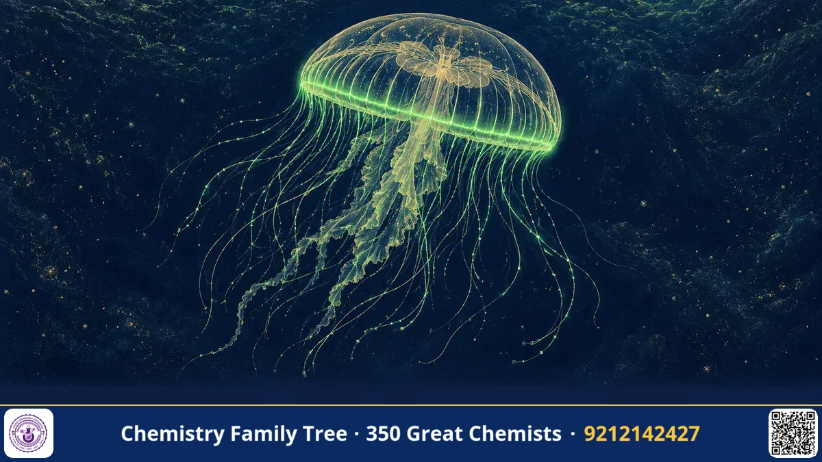
Today scientists can watch cancer cells spread, nerves wire a brain or bacteria infect a plant by making them glow green. The tool behind these films is green fluorescent protein (GFP), a protein from the crystal jellyfish Aequorea victoria. It was first isolated and characterised by the Japanese chemist Osamu Shimomura in the early 1960s, when he was trying to find out why the jellyfish gives off light. In 2008 he shared the Nobel Prize in Chemistry with Martin Chalfie and Roger Tsien "for the discovery and development of the green fluorescent protein, GFP".
The Scientific Problem Before the Discovery
Living organisms produce light in a process called bioluminescence, as in fireflies, deep-sea fish and some marine animals. Chemically, it is a reaction in which a substrate (a luciferin) is oxidised by an enzyme (a luciferase), and part of the energy is released as visible light rather than as heat. By the 1950s several luciferins were known, and Shimomura, then a young researcher at Nagoya University, had already succeeded in crystallising luciferin from the small sea crustacean Cypridina and determining its structure, which was considered an almost impossible task.
The glowing jellyfish Aequorea posed a new puzzle. Its light came from the rim of the bell, but the colour of the light seen in the animal was green, whereas extracts of the light-producing material gave blue light. Why the difference? And what was the trigger?
The Discovery: Step by Step
- Collecting jellyfish (1961): Shimomura travelled with his mentor Frank Johnson from Princeton University to the Friday Harbor Laboratories in Washington State. They collected jellyfish from the docks in thousands during the summer. Over the years tens of thousands of animals were handled.
- Extraction: The glowing rings were cut off, squeezed and the extract was purified by column chromatography and other protein separation methods.
- The accidental clue: A well-known story is that Shimomura poured extract into a sink, and the sink glowed brightly. Seawater, containing Ca²⁺, had triggered the light. The extract needed calcium ions to emit light.
- Aequorin (1962): The team purified a protein that emitted blue light when Ca²⁺ was added. They named it aequorin. It did not need oxygen added from outside, since the oxygen was already bound to the protein-luciferin complex.
- GFP: During the purification they noticed another protein which was not luminescent but fluoresced green under ultraviolet light. This was the green fluorescent protein. It absorbed the blue light from aequorin (about 470 nm) and re-emitted it as green light (about 509 nm), explaining the colour in the living animal.
- Chromophore structure (1979): Shimomura worked out that the light-absorbing group of GFP is formed from three amino-acid residues in the protein chain, and proposed its structure.
The Chemistry Behind It
The light reaction of aequorin involves the luciferin coelenterazine bound inside the protein as a peroxide:
Aequorin (coelenterazine-OO⁻) + Ca²⁺ → coelenteramide* + CO₂ → coelenteramide + hν (blue, ~470 nm)
Binding of Ca²⁺ to the protein changes its shape. The coelenterazine hydroperoxide undergoes cyclisation to a dioxetanone, which breaks down with loss of CO₂, forming the excited-state amide. When it relaxes, a photon of blue light is emitted. Within the jellyfish, this energy is passed to GFP without emitting a photon (Förster resonance energy transfer), and the green protein emits its characteristic light.
GFP is a beautiful example of structural chemistry. It has 238 amino acids and its shape is a "β-barrel" of eleven β-strands enclosing a central α-helix, like a can. The chromophore sits at the centre of the barrel, shielded from water and quenching. It is formed spontaneously, by a post-translational process requiring only molecular oxygen:
Ser65–Tyr66–Gly67 → cyclisation (imidazolinone ring) → oxidation by O₂ (–2H) → conjugated p-hydroxybenzylidene-imidazolinone chromophore
The extended conjugation lowers the energy gap so that the chromophore absorbs near 395 nm (and 475 nm in the anionic state) and fluoresces green. No cofactor or enzyme is required, which is why GFP can be expressed in any organism: Martin Chalfie showed in 1994 that it glows in bacteria and in roundworm neurons, and Roger Tsien made variants of other colours.
Applications Today
- Cell biology: Fusing GFP to a protein of interest lets scientists watch where it goes in living cells.
- Gene expression reporters: GFP is used to show when and where a gene is switched on.
- Neuroscience: Colourful "Brainbow" mice and tracing of nerve connections.
- Medicine: Tracking tumour cells, bacteria and viruses; measuring calcium with aequorin-based sensors.
- Agriculture and environment: GFP-tagged bacteria help to study soil microbes and biosensors for arsenic or pollutants.
Exam Corner
- Shimomura shared the Nobel Prize in Chemistry 2008 with Martin Chalfie and Roger Y. Tsien.
- Aequorin emits blue light (~470 nm) on binding Ca²⁺; GFP emits green light (~509 nm).
- GFP: 238 amino acids, β-barrel, chromophore from Ser65-Tyr66-Gly67 formed by cyclisation and oxidation.
- Bioluminescence is chemiluminescence in a living organism: luciferin + O₂ → oxyluciferin* → light.
- Fluorescence involves absorption at a short wavelength and emission at a longer one (Stokes shift).
Common Misconceptions
- "GFP produces light by itself." It does not; it fluoresces after absorbing light, either from UV/blue illumination or from aequorin.
- "Shimomura engineered GFP in other organisms." That was Chalfie and Tsien; Shimomura discovered and characterised the protein and its chromophore.
- "Fluorescence and bioluminescence are the same." In fluorescence, light is absorbed first; in bioluminescence, a chemical reaction creates the excited state.
Quick Revision Box
- Source: jellyfish Aequorea victoria; work done at Friday Harbor, 1961 onwards.
- Aequorin + Ca²⁺ gives blue light; GFP shifts it to green.
- GFP chromophore: cyclised Ser-Tyr-Gly, oxidised by O₂.
- Structure: 11-strand β-barrel around a central helix.
- Nobel Prize in Chemistry 2008: Shimomura, Chalfie, Tsien.
- Uses: a genetically encoded fluorescent tag.
FAQ
Why does the jellyfish need GFP?
The biological purpose is not fully clear, but GFP converts the blue light of aequorin into green light, which is how the jellyfish appears in the sea.
Why is GFP so useful?
It forms its chromophore by itself, needs only oxygen, and is non-toxic, so it can be expressed in nearly any cell.
What does Ca²⁺ do in aequorin?
It binds to the protein, triggering the breakdown of the bound luciferin peroxide and the emission of light, which makes aequorin a calcium indicator.
Was it a Nobel Prize in Medicine?
No, it was awarded in Chemistry.
Published by ABC Chemistry — IIT-JAM | CSIR-NET | GATE Chemistry Coaching. Visit abcchemistry.com
About the chemist
Osamu Shimomura
1928-2018 · Japan · Nobel Chemistry 2008
Discovery of green fluorescent protein