The discovery, explained · Chemist no. 321 of 350
James Watson, Francis Crick and the DNA Double Helix
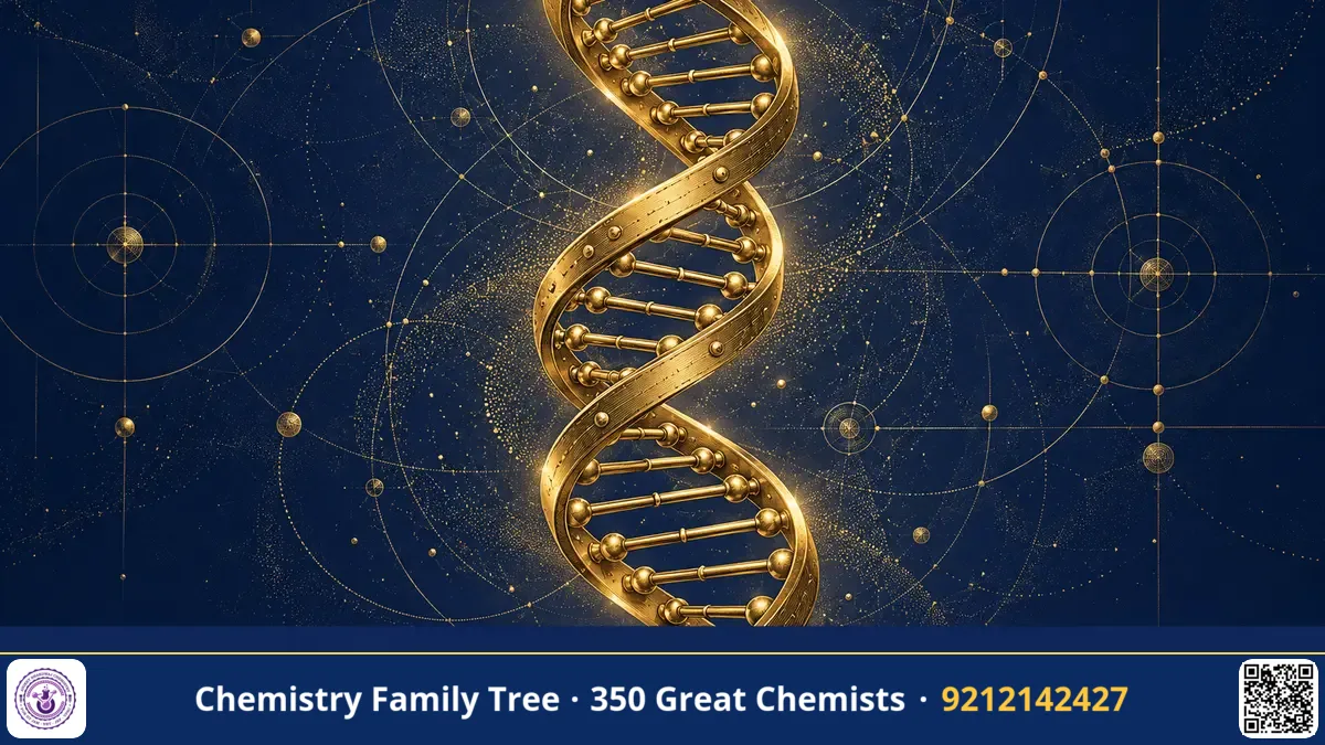
On 25 April 1953 the journal Nature published a one-page paper that changed biology forever. In it, James Watson (born 1928) and Francis Crick (1916-2004) proposed that DNA is a double helix: two chains wound around each other, held together by pairs of bases. They wrote one of the most famous understatements in science: "It has not escaped our notice that the specific pairing we have postulated immediately suggests a possible copying mechanism for the genetic material." The structure explained how genes can be copied and how information is stored. Watson, Crick and Maurice Wilkins shared the 1962 Nobel Prize in Physiology or Medicine. The work rested on the X-ray diffraction data from Rosalind Franklin and Wilkins, and on the chemical rules of Erwin Chargaff.
The Scientific Problem Before the Discovery
By the early 1950s, evidence was mounting that DNA, not protein, carries heredity. Avery, MacLeod and McCarty (1944) showed that DNA transformed bacteria, and Hershey and Chase (1952) confirmed it with radioactive phosphorus (32P) in DNA and sulphur (35S) in protein of bacteriophage. But what did DNA look like? How could a molecule made of only four kinds of units store the instructions for a whole organism, and how could it be copied exactly each time a cell divides?
Chemists knew the parts: a sugar (deoxyribose), a phosphate group and four bases, adenine (A), guanine (G), cytosine (C) and thymine (T), linked in chains by 3'-5' phosphodiester bonds (Todd and others). Linus Pauling had just shown that proteins can form an α-helix, and he was working on a DNA model, a triple helix with the phosphates inside, which turned out to be wrong.
The Discovery / Experiment
Step 1: Chargaff's rules
Erwin Chargaff analysed DNA from many species by paper chromatography and found that in every DNA the amount of A equals T and the amount of G equals C, even though the ratio (A+T)/(G+C) varies from species to species. Crick and Watson later realised that this one-to-one ratio hints at pairing.
Step 2: X-ray diffraction
At King's College London, Maurice Wilkins and Rosalind Franklin (with her student Raymond Gosling) produced X-ray diffraction patterns of DNA fibres. Franklin showed that DNA exists in two forms, a dry A form and a wet B form. Her B-form picture, called Photo 51 (taken in May 1952), showed a clear X-shaped pattern, the typical sign of a helix. From it one can calculate the distance between repeating units: the helix repeats every 34 Å, the bases are spaced 3.4 Å apart (so 10 base pairs per turn), and the diameter is about 20 Å. Watson saw the photograph, shown to him by Wilkins, in January 1953, and Crick saw Franklin's report data via Max Perutz. This sharing of data happened without Franklin's knowledge, which is an important ethical point in the history of this discovery.
Step 3: Model building in Cambridge
At the Cavendish Laboratory, Watson and Crick built physical models using metal plates and rods, like a three-dimensional jigsaw. A first model with a three-chain structure and the phosphates inside was a failure. After seeing Franklin's data they put the sugar-phosphate backbones on the outside, and the bases on the inside. The bases could not be placed at random: purines (A, G) are two-ringed and pyrimidines (T, C) are single-ringed, so a purine paired with a pyrimidine would give a uniform width of 20 Å.
Step 4: The correct base pairs
An important point came from Jerry Donohue, a chemist in their office, who told Watson that the bases exist mainly in the keto (lactam) form and not in the enol form given in textbooks. With the keto forms, the pairs fitted perfectly:
A = T   (two hydrogen bonds)
G ≡ C   (three hydrogen bonds)
Both pairs have nearly the same geometric shape, so any sequence of bases can fit into the helix. The two chains run in opposite directions (antiparallel): one 5' to 3', the other 3' to 5'.
Step 5: Implications
Because A always pairs with T, and G with C, each strand determines the other. If the strands are separated, each can act as a template to build a new complementary strand. This is semi-conservative replication, which was confirmed by Meselson and Stahl in 1958 with 15N-labelled bacteria and density gradient centrifugation.
The Chemistry Behind It
- Backbone: alternating deoxyribose and phosphate groups joined through 3'-5' phosphodiester bonds; each phosphate carries a negative charge, which is why DNA is an acid and binds to positively charged histone proteins.
- Hydrogen bonding: A–T: two H-bonds, G–C: three, so GC-rich DNA is more stable and has a higher melting temperature (Tm).
- Stacking: π–π stacking between bases and hydrophobic effects give most of the stability of the helix, more than the hydrogen bonds themselves.
- Grooves: The helix has a wide major groove and a narrow minor groove, where proteins recognise base sequences.
- Forms: B-DNA (right-handed, 10 bp per turn, the physiological form), A-DNA (right-handed, 11 bp per turn, dehydrated) and Z-DNA (left-handed, zig-zag, with alternating purine-pyrimidine sequences).
- Chargaff: A = T and G = C, hence purines = pyrimidines.
Applications Today
- DNA fingerprinting and forensics: Used in Indian courts and by CFSL laboratories for criminal and paternity cases.
- PCR and diagnostics: Primer annealing depends on Watson-Crick base pairing.
- Genome sequencing: The Human Genome Project (1990-2003), in which Watson was the first director, and India's GenomeIndia project.
- Drug design: Many anticancer drugs, such as cisplatin, doxorubicin, and DNA intercalators, bind DNA.
- Gene editing and mRNA vaccines: CRISPR guide RNAs find targets by base pairing.
Exam Corner
- Watson-Crick model (1953): right-handed, antiparallel double helix, 20 Å diameter, 34 Å pitch, 3.4 Å rise, 10 bp per turn (B-DNA).
- Chargaff's rules: A = T, G = C; (A+T)/(G+C) is species-specific.
- Nobel Prize 1962, Physiology or Medicine, to Watson, Crick and Wilkins.
- Melting temperature rises with GC content (three H-bonds).
- Semi-conservative replication proved by Meselson and Stahl using 15N.
Common Misconceptions
- "Watson and Crick discovered DNA." No. DNA was isolated by Friedrich Miescher in 1869; they discovered its three-dimensional structure.
- "Hydrogen bonds alone hold the helix together." Base stacking contributes more to stability.
- "The Nobel Prize was in Chemistry." It was in Physiology or Medicine. Rosalind Franklin had died in 1958, and the Nobel Prize is not awarded posthumously.
Quick Revision Box
- Published in Nature, 25 April 1953.
- Two antiparallel strands, sugar-phosphate backbone outside, bases inside.
- A–T (2 H-bonds), G–C (3 H-bonds); purine pairs with pyrimidine.
- B-DNA: 10 bp per turn, 34 Å pitch, 20 Å diameter.
- Key data: Franklin and Wilkins X-ray, Chargaff's rules, Donohue's keto forms.
- Nobel Prize 1962, Physiology or Medicine.
FAQ
Why did the base pairs have to be purine-pyrimidine?
Two purines would be too wide and two pyrimidines too narrow for the constant 20 Å width of the helix seen in X-ray patterns.
What was Photo 51?
It was an X-ray diffraction picture of B-DNA taken by Rosalind Franklin and Raymond Gosling in 1952, showing the cross pattern of a helix.
Who got the Nobel Prize for the double helix?
James Watson, Francis Crick and Maurice Wilkins in 1962, in Physiology or Medicine. Franklin had died in 1958.
Why is the double helix so important for exams?
It is the base for questions on replication, transcription, melting temperature, Chargaff's rules, DNA forms and genetic engineering in all competitive exams.
Published by ABC Chemistry — IIT-JAM | CSIR-NET | GATE Chemistry Coaching. Visit abcchemistry.com
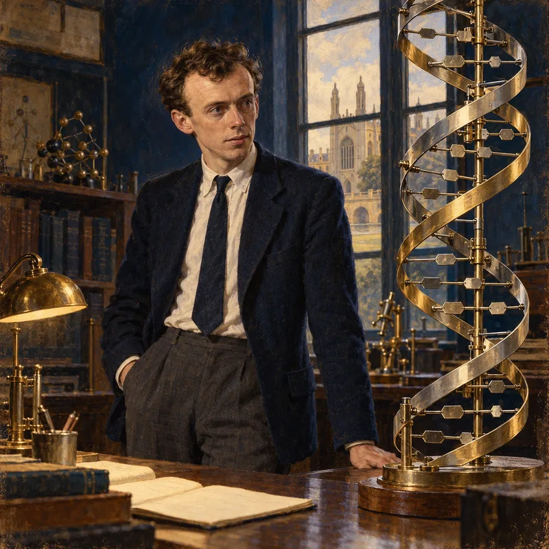
About the chemist
James Watson
1928-2025 · United States · Nobel Physiology/Medicine 1962
Co-discovered DNA double helix