The discovery, explained · Chemist no. 125 of 350
Arne Tiselius: How Electric Fields Taught Us to See Blood Proteins
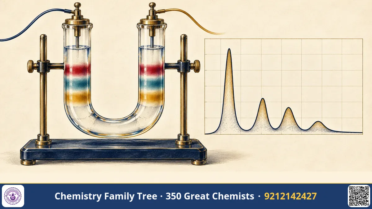
When a doctor orders a "serum protein electrophoresis" test, the report shows a curve with a tall peak for albumin, small peaks for alpha and beta globulins, and a broad peak for gamma globulins. This test is a direct descendant of the work of the Swedish biochemist Arne Wilhelm Kaurin Tiselius. In the 1930s he built an apparatus that could separate proteins in an electric field and used it to show that blood serum is a complex mixture, not a single "protein". He also advanced adsorption chromatography. For "his research on electrophoresis and adsorption analysis, especially for his discoveries concerning the complex nature of the serum proteins", he received the Nobel Prize in Chemistry for 1948.
The Scientific Problem Before the Discovery
By 1930 chemists understood that proteins are large molecules made of amino acids, and that they carry electric charge in solution. The amino and carboxyl groups of the side chains ionise depending on the pH. Each protein has an isoelectric point (pI), the pH at which its net charge is zero. Below the pI it is positively charged, and above the pI negatively charged.
- Proteins from blood, egg or milk were purified by precipitation with salts or alcohol, but chemists did not know how pure the fractions were.
- Earlier work on moving boundaries in an electric field existed, but the results were not reliable because of convection currents and heating.
- Serum was thought to contain mainly albumin and "globulin", and the true number of components, and their roles in immunity, were unknown.
The Discovery: Step by Step
The moving-boundary electrophoresis apparatus
Tiselius, as a doctoral student of The Svedberg at Uppsala, started with Svedberg's ultracentrifuge ideas and designed a new instrument to measure how fast proteins move in an electric field. His doctoral thesis (1930) described the method, and in 1937 he published an improved apparatus that became the "Tiselius apparatus".
- The cell. A U-shaped glass tube, made of sections that can slide against each other, was filled with a buffer in the upper parts and a protein solution in the bottom of the U.
- Cooling. The cell was placed in a water bath at about 4 °C, where water has its maximum density. This reduced convection from the heat produced by the current, which had ruined earlier experiments.
- Electric field. Electrodes at the ends of the U-tube applied a voltage. Positively charged proteins moved to the cathode, and negative ones moved to the anode, each type at its own speed.
- Optical detection. Boundaries between buffer and protein solution were seen by the change of refractive index, using a Schlieren optical system (invented by Toepler) that shows gradients of concentration as peaks.
- Analysis. The positions of the peaks over time gave the mobility of each component. The area under each peak was proportional to concentration.
The mobility (μ) is the velocity per unit field strength:
μ = v / E     (units: cm² V⁻¹ s⁻¹)
For a sphere of charge Q and radius r in a liquid of viscosity η:  μ ≈ Q / (6πηr)
The complex nature of serum
Using the apparatus, Tiselius showed in 1937 that human and animal serum, when run at pH 8 in a buffer, gives five main peaks: albumin (fastest toward the anode), then α₁-, α₂-, β- and γ-globulins (the slowest). Albumin is the main protein of blood and helps maintain osmotic pressure and carries fatty acids and drugs. The α- and β-globulins include transport proteins, such as transferrin and lipoproteins. The γ-globulins attracted special attention.
In 1939, Tiselius and the American immunologist Elvin Kabat showed that when a rabbit is immunised, the antibodies appear in the γ-globulin fraction. After immunisation, the γ-peak became larger, and the antibody activity vanished from the serum when the antigen removed the antibodies, with the γ peak falling. This was the first physical proof that antibodies are proteins in a specific fraction of serum. Later, the study of the γ-globulin fraction showed its change in diseases: in multiple myeloma a sharp, narrow band appears in the electrophoretic pattern, while in immune deficiency the γ region is low.
Adsorption analysis and chromatography
Tiselius also improved the chromatography first used by Tswett. He introduced systematic methods in the late 1930s and 1940s, including frontal analysis and displacement development, and developed ways to analyse colourless substances, using the Schlieren optical system. He showed that calcium phosphate (hydroxyapatite) can be used as an adsorbent for proteins. His group laid the groundwork for ion exchange and gel filtration methods. His students Jerker Porath and Per Flodin developed gel filtration (Sephadex) in 1959, and Stellan Hjertén worked on capillary electrophoresis and later forms of chromatography.
The Chemistry Behind It
Charge and pH. A protein with an isoelectric point of 5 is negative at pH 8 and migrates toward the anode. Albumin (pI near 4.7) has a high negative charge at pH 8, so it migrates fastest. The γ-globulins have the highest pI of the serum fractions, so they carry the smallest net negative charge at pH 8 and travel the least.
Buffers and ionic strength. A buffer is needed to fix the pH and to carry the current. High ionic strength reduces the mobility, because the counter-ion atmosphere shields the protein. Therefore ionic strength must be kept low and constant. The heat produced is I²R, and thus the current must not be too great.
Adsorption. A molecule in solution is in equilibrium with the surface of an adsorbent. Strongly adsorbed molecules move slowly through a column and weakly adsorbed ones move fast, which separates them. Adsorption isotherms, such as the Langmuir isotherm, describe this:
θ = K·c / (1 + K·c)
Applications Today
- Clinical diagnosis. Serum protein electrophoresis helps to detect multiple myeloma, chronic inflammation, liver disease, nephrotic syndrome and immunodeficiency. It is in every pathology laboratory in India.
- Haemoglobin tests. Electrophoresis (and HPLC) detect abnormal haemoglobins, such as HbS in sickle cell disease and HbE and β-thalassaemia, which are important in India.
- Molecular biology. Gel electrophoresis of DNA, RNA and proteins is a daily tool. Agarose gels separate DNA fragments in PCR and forensic DNA profiling, and SDS-PAGE separates proteins by size.
- Chromatography. Ion exchange, affinity and size exclusion chromatography are used in making pharmaceuticals such as insulin and antibodies.
- Genome sequencing. Capillary electrophoresis was the heart of the Sanger-based sequencing machines used in the Human Genome Project.
Exam Corner
- Tiselius built the moving-boundary electrophoresis apparatus (1937) and showed five serum fractions: albumin, α₁, α₂, β and γ-globulins.
- Electrophoretic mobility μ = v/E; it depends on charge, size and shape, and on pH and ionic strength.
- Isoelectric point: pH at which the net charge of a protein (zwitterion) is zero; the mobility is zero.
- Antibodies are found in the γ-globulin fraction (Tiselius and Kabat, 1939).
- Nobel Prize in Chemistry 1948 for electrophoresis and adsorption analysis.
Common Misconceptions
- "Proteins migrate in an electric field because they are always negatively charged." The charge depends on the pH relative to the pI.
- "Electrophoresis separates only by charge." In free solution it depends on charge-to-size ratio. In gels, sieving by size also matters.
- "Tiselius invented gel electrophoresis." His method was moving-boundary electrophoresis in free solution. Gel and zone electrophoresis developed later.
Quick Revision Box
- Tiselius apparatus: U-tube, cooled to about 4 °C, Schlieren optics.
- Serum peaks: albumin, α₁, α₂, β, γ.
- Albumin is the fastest toward the anode at pH 8.
- Antibodies are in γ-globulin (1939, with Kabat).
- Student work led to gel filtration (Porath, Flodin).
- Nobel Prize in Chemistry, 1948.
FAQ
Why did Tiselius cool the cell to 4 °C?
Water is at its densest near 4 °C, so heating by the current produces the smallest convection currents, and the boundaries stay sharp.
What is the difference between electrophoresis and chromatography?
Electrophoresis separates charged molecules moving in an electric field. Chromatography separates molecules by differing distribution between a stationary and a mobile phase.
Which serum protein is the most abundant?
Albumin, which makes about 55-60% of serum protein.
Was the Nobel Prize in Medicine?
No. It was the Nobel Prize in Chemistry for 1948.
Published by ABC Chemistry — IIT-JAM | CSIR-NET | GATE Chemistry Coaching. Visit abcchemistry.com
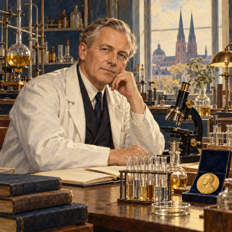
About the chemist
Arne Tiselius
1902-1971 · Sweden · Nobel Chemistry 1948
Electrophoresis and adsorption analysis