The discovery, explained · Chemist no. 196 of 350
Brian Kobilka and the First Look at a Working GPCR
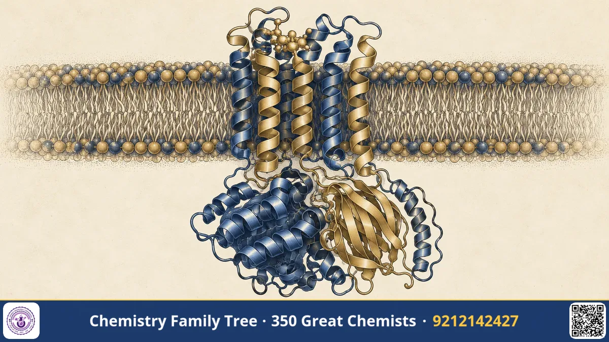
Receptors on the surface of your cells are the "ears" through which hormones and neurotransmitters speak. Robert Lefkowitz had shown that the beta-adrenergic receptor exists and had cloned its gene. But the picture was still a sequence on paper. Brian K. Kobilka, who began as Lefkowitz's postdoctoral fellow and later led his own laboratory at Stanford University, wanted to see the receptor in three dimensions. In 2007 his group solved the first crystal structure of a human GPCR bound to a drug, and in 2011 they captured the receptor in its active state, bound to a G protein. He shared the 2012 Nobel Prize in Chemistry with Lefkowitz. This post explains how this "impossible" structure was achieved and why it matters.
The Scientific Problem Before the Discovery
By the late 1980s it was clear that GPCRs formed a huge family with a common seven-transmembrane helix design. Only one such structure was known at atomic detail: bovine rhodopsin, solved by Krzysztof Palczewski and co-workers in 2000. Rhodopsin is unusual because it is abundant in the retina and very stable. Hormone and neurotransmitter receptors, in contrast, are present in tiny quantities and are unstable once removed from the membrane.
Three barriers stood in the way of crystallography. First, GPCRs are hydrophobic membrane proteins, and must be extracted with detergents that can strip away the lipids they need. Second, they are very flexible: the receptor flips between inactive and active forms, and a flexible protein resists forming an ordered crystal. Third, producing enough protein, in a pure and homogeneous form, was difficult. Many leading laboratories tried and failed for years.
The Discovery: Step by Step
- Cloning and biochemistry (1980s). At Duke, Kobilka helped clone the gene for the β₂-adrenergic receptor and then explored which regions determine ligand binding and G protein coupling, for example by swapping segments between different receptors to build chimeras.
- Producing large amounts. At Stanford he developed ways to express the receptor in insect cells and purify it in detergent. His group also used fluorescence spectroscopy to watch how the receptor changes shape when ligands bind, showing it is a dynamic molecule that explores several conformations.
- Stabilising the receptor. The team used a ligand with strong, long-lasting binding, which freezes the receptor in one state, and replaced the flexible third intracellular loop with T4 lysozyme, a small, well-behaved protein that makes crystal contacts. They also grew crystals in a lipid mesophase (lipidic cubic phase) with the help of Raymond Stevens' group, which keeps the receptor in a membrane-like environment.
- The 2007 structure. The first high-resolution crystal structure of a human β₂-adrenergic receptor bound to an inverse agonist (carazolol) was published in 2007, in a collaboration between several laboratories.
- The active complex (2011). The group then captured the β₂ receptor bound to an agonist and to the stimulatory G protein Gs, stabilised using a nanobody (a small antibody fragment from camelids) with collaborators in several laboratories. The crystal structure, published in 2011, showed for the first time how a receptor activates a G protein.
The Chemistry Behind It
The β₂ receptor has seven α-helices (TM1-TM7) forming a bundle, with the ligand-binding pocket buried among them. When an agonist such as adrenaline binds, small movements at the extracellular side are amplified, and the cytoplasmic end of TM6 swings outward by roughly 14 Å. This opens a cavity for the α5 helix of the G protein's Gα subunit to insert. In Gα this causes the nucleotide-binding domain to open, GDP is released, and GTP (which is more abundant in the cell) binds:
Receptor* + Gα·GDP·βγ → Receptor*·Gα(empty)·βγ → Receptor* + Gα·GTP + βγ
Binding of ligands to the pocket involves hydrogen bonds (for example, between the catechol hydroxyl groups of adrenaline and serine residues in TM5), an ionic interaction between the protonated amine and an aspartate (Asp113 in TM3), and aromatic stacking with phenylalanine residues. The ligand categories are:
| Ligand type | Effect on receptor | Example |
|---|---|---|
| Full agonist | Strongly stabilises active state | Adrenaline, isoprenaline |
| Partial agonist | Weaker activation | Some beta agonists |
| Antagonist | Blocks agonist binding | Propranolol (non-selective beta-blocker) |
| Inverse agonist | Reduces basal activity | Carazolol |
The structures also taught that GPCRs are "molecular switches": the equilibrium between inactive (R) and active (R*) states is shifted by ligands, as in the allosteric two-state model.
Applications Today
- Structure-based drug design. Companies can dock candidate molecules into receptor pockets on a computer, saving years in drug discovery.
- Better beta drugs. Understanding why some beta-blockers are selective for β₁ (heart) and others for β₂ (lungs) helps avoid side effects such as bronchospasm.
- Many other targets. Methods developed for the β₂ receptor are used on opioid, dopamine, serotonin and muscarinic receptors, among others.
- Cryo-EM era. The G protein complex gave a template for modern cryo-electron microscopy structures of GPCR-G protein assemblies.
- Safer pain-relief research. Studying how opioid receptors couple to G proteins versus arrestins may lead to better analgesics.
Exam Corner
- Nobel Prize in Chemistry 2012: Lefkowitz and Kobilka, for studies of GPCRs.
- First human GPCR (β₂-AR) crystal structure: 2007; active state with Gs: 2011. Bovine rhodopsin had been solved in 2000.
- Strategy: T4 lysozyme fusion replacing intracellular loop 3, lipidic cubic phase and a nanobody to stabilise the active state.
- Activation: outward movement of TM6 opens a binding site for the Gα C-terminal helix; GDP is exchanged for GTP.
- Agonist, antagonist, inverse agonist: know the difference.
Common Misconceptions
- "GPCRs are rigid locks and ligands are keys." They are flexible and exist as an ensemble of states; ligands shift the balance.
- "Rhodopsin is a typical GPCR in terms of stability." It is unusually abundant and stable; most other GPCRs are not.
- "G proteins are the same as small GTPases like Ras." The Gα subunit is a GTPase, but heterotrimeric G proteins are different from monomeric Ras-type proteins.
Quick Revision Box
- Kobilka: postdoc with Lefkowitz, later Stanford professor.
- 2007: structure of the human β₂-adrenergic receptor.
- 2011: β₂-AR–Gs complex (agonist-bound, nanobody-stabilised).
- Tools: T4 lysozyme fusion, lipidic cubic phase, nanobody.
- Activation: TM6 moves outward by about 14 Å.
- Nobel Prize in Chemistry 2012.
FAQ
What is a nanobody?
A nanobody is a small antibody fragment derived from camelids such as llamas. Its small size and stability make it a useful tool for locking proteins in specific conformations.
Why replace a loop with T4 lysozyme?
The loop is flexible and prevents crystal packing. T4 lysozyme is rigid and crystallises well, so it creates contacts between receptor molecules in the lattice.
What is lipidic cubic phase?
It is a gel-like lipid matrix with channels, in which membrane proteins can diffuse and organise into crystals in a more natural environment than detergent alone.
Is this relevant to pharmaceutical chemistry exams?
Yes. Receptor structure, ligand types and binding interactions are core drug-design topics.
Published by ABC Chemistry — IIT-JAM | CSIR-NET | GATE Chemistry Coaching. Visit abcchemistry.com
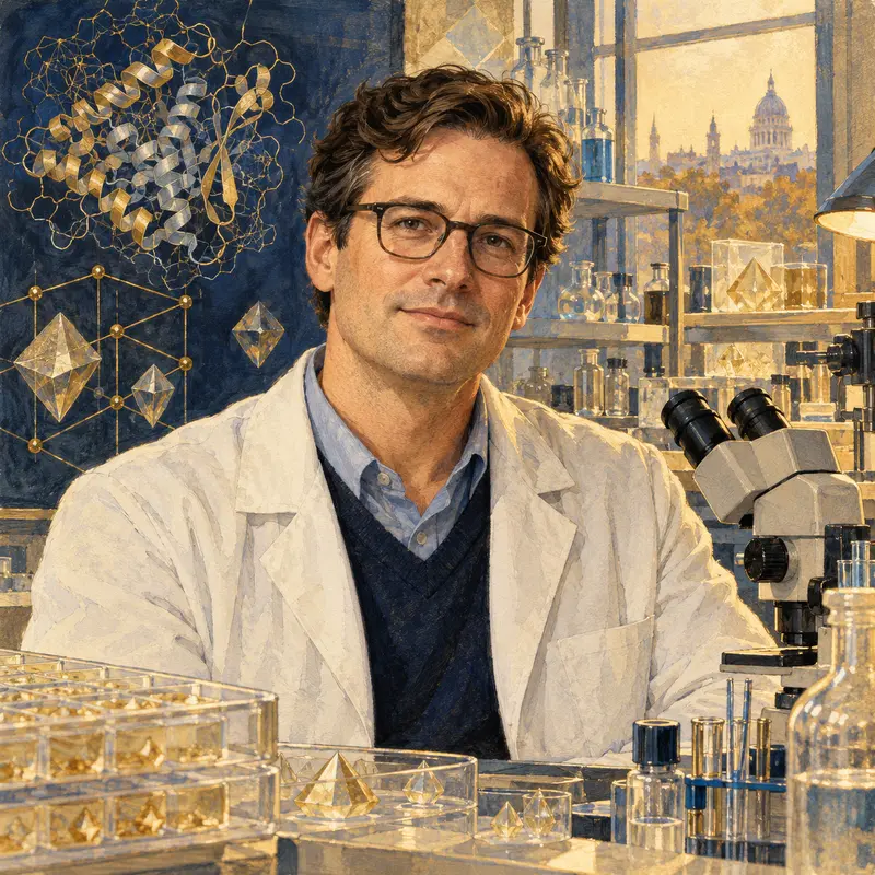
About the chemist
Brian Kobilka
1955- · United States · Nobel Chemistry 2012
Structure of G-protein-coupled receptors