The discovery, explained · Chemist no. 66 of 350
Dorothy Hodgkin: How X-ray Crystallography Revealed Penicillin, Vitamin B12 and Insulin
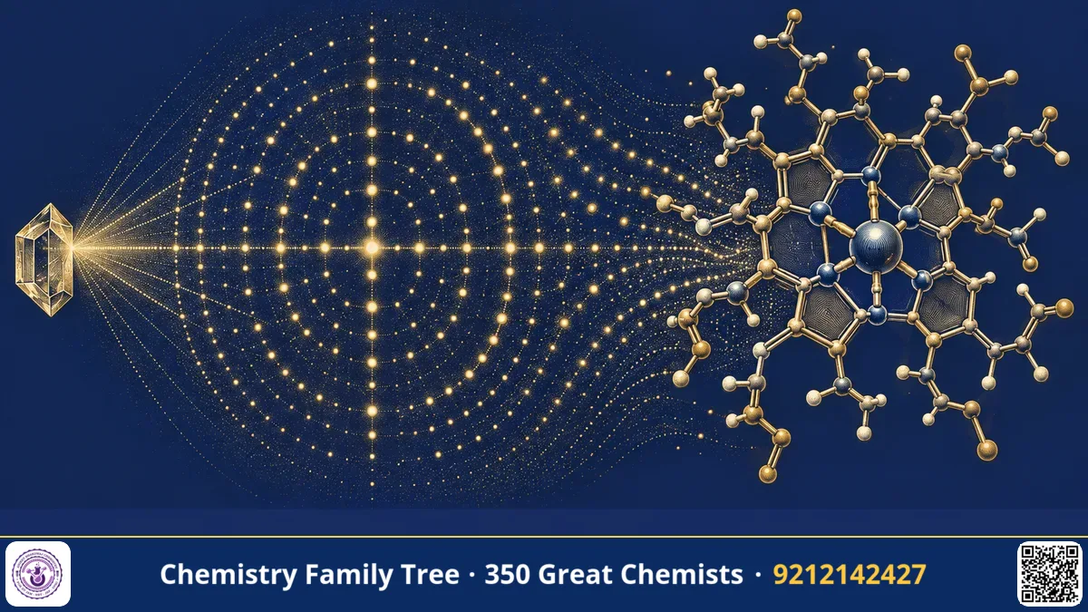
Imagine trying to draw a molecule that is far too small to see under any microscope. You can only shine X-rays on a crystal of it, record a pattern of spots on a photographic plate, and then work backwards to find where every atom sits. This is exactly what Dorothy Crowfoot Hodgkin did, and she did it for some of the most important molecules in medicine: cholesterol-related compounds, penicillin, vitamin B12 and insulin. For this work she won the Nobel Prize in Chemistry in 1964. Her results proved that X-ray methods could solve structures of real biological importance and opened the path to modern structural biology and drug design. In this post we follow her discoveries step by step, learn the chemistry behind them, and collect the points that matter for IIT-JAM, CSIR-NET, GATE and board exams.
The Scientific Problem Before the Discovery
In the 1930s chemists decided a structure by degradation and synthesis. You broke a molecule into smaller pieces, identified the pieces, and then guessed how they were joined. This worked well for small molecules but failed badly for complex natural products. Penicillin was a famous example. During the Second World War the drug was urgently needed, yet its structure was disputed. Some chemists proposed a thiazolidine fused to an oxazolone ring, while others believed it contained a strained four-membered β-lactam ring. Chemical evidence alone could not settle the matter.
Vitamin B12 was an even bigger puzzle. It was known to cure pernicious anaemia, but it was a huge molecule containing cobalt, phosphorus, nitrogen and a very unusual ring system. Degradation gave fragments that nobody could assemble into a single picture. What chemistry needed was a method that could look at the whole molecule at once.
The Discovery: Step-by-Step Structure Solving
Hodgkin trained in crystallography at Cambridge under J. D. Bernal, who had taken some of the first X-ray photographs of a protein crystal (pepsin) in 1934. She then built her career at Oxford. Her general method followed these steps:
- Grow a good crystal. The molecule must pack in a regular lattice. This is often the hardest step.
- Record the diffraction pattern. X-rays are scattered by electrons, and the scattered waves reinforce only in certain directions, giving spots on film.
- Measure spot positions and intensities. Positions give the unit cell size; intensities carry information about atom positions.
- Solve the phase problem. Detectors record intensity but not phase. Hodgkin used heavy atoms (for example bromine or iodine, or the cobalt already present in B12) because they scatter strongly and help fix phases.
- Calculate an electron density map by Fourier synthesis and fit the atoms into it.
For penicillin, Hodgkin and her colleagues worked on crystalline salts of the drug during the war years. Their analysis showed the fused β-lactam and thiazolidine ring system, settling the argument. The result was published in 1949.
For vitamin B12 (formula C63H88CoN14O14P), she collected thousands of reflections and used early electronic computers to handle the heavy Fourier calculations. In 1956 she reported the full structure: a cobalt atom at the centre of a corrin ring, which is like a porphyrin but with one direct link between two pyrrole rings, with a dimethylbenzimidazole nucleotide attached below. This was the first time a molecule of such complexity had been solved purely by X-ray analysis.
For insulin she had photographed the first crystals in 1934, but the structure needed decades more of work. She finally reported the three-dimensional structure in 1969 after about 35 years of effort.
The Chemistry Behind It
The central equation of the method is Bragg's law:
nλ = 2d sinθ
Here λ is the X-ray wavelength, d is the spacing between lattice planes, θ is the angle of incidence and n is the order of reflection. Because atoms in a molecule are about 1 to 1.5 Å apart, X-rays with wavelength near 1.5 Å (for example Cu Kα) are suitable.
The scattered amplitude from a plane is called the structure factor, F(hkl). The electron density at a point (x, y, z) is obtained by a Fourier sum over all reflections:
ρ(x,y,z) = (1/V) Σ F(hkl) exp[-2πi(hx + ky + lz)]
Since only |F|² is measured, phases are missing, and this is the phase problem. The heavy-atom method works because a single heavy atom dominates scattering, so its position can be found first (using a Patterson map) and its phases used as a starting estimate.
The chemistry results are also valuable. The β-lactam ring of penicillin is a four-membered cyclic amide with high ring strain, so it acylates the bacterial transpeptidase enzyme that builds the cell wall. In B12, the Co–C bond in the coenzyme form is one of the very few natural organometallic bonds, and the cobalt cycles between Co(I), Co(II) and Co(III).
Applications Today
- Antibiotics: Knowing the β-lactam core allowed chemists to make semi-synthetic penicillins such as ampicillin and amoxicillin, which are widely used in India.
- Nutrition and medicine: B12 deficiency is common among vegetarians, and treatment uses injections or tablets of cyanocobalamin or methylcobalamin.
- Diabetes care: Insulin structure guided the design of fast-acting and long-acting analogues.
- Drug design: Pharmaceutical companies routinely solve protein-ligand crystal structures to design inhibitors. Synchrotron facilities, including India's Indus-2 at Indore, serve crystallographers.
Exam Corner
- Write Bragg's law and be ready to find d or θ from given values of λ and n.
- X-rays are scattered by electrons, so hydrogen atoms are hard to locate; neutron diffraction scatters from nuclei.
- Vitamin B12 contains cobalt in a corrin ring; the corrin differs from porphyrin by lacking one meso carbon bridge.
- Penicillin contains a β-lactam fused to a thiazolidine ring; the strained four-membered ring is responsible for activity.
- Hodgkin won the 1964 Nobel Prize in Chemistry alone; she is the third woman to win it after Marie Curie and Irène Joliot-Curie.
Common Misconceptions
- "X-rays take a photograph of a molecule." No. The pattern is a diffraction pattern, and the image is reconstructed mathematically.
- "Hodgkin discovered penicillin." Alexander Fleming discovered it in 1928; Florey and Chain developed it as a drug. Hodgkin determined its structure.
- "Vitamin B12 is a metal-free vitamin." It is one of the few biomolecules that contain cobalt.
Quick Revision Box
- Method: single-crystal X-ray diffraction, Bragg's law nλ = 2d sinθ.
- Penicillin structure confirmed (published 1949): β-lactam plus thiazolidine ring.
- Vitamin B12 structure solved 1956: Co(III) in a corrin ring.
- Insulin structure reported 1969 after about 35 years.
- Phase problem solved using heavy atoms.
- Nobel Prize in Chemistry 1964.
FAQ
Why did Hodgkin use heavy atoms?
Heavy atoms scatter X-rays strongly. Their positions can be located easily, and they provide approximate phases for the whole structure.
What is a corrin ring?
A corrin is a macrocycle of four reduced pyrrole-type rings similar to a porphyrin, but two of the rings are joined directly without a methine bridge. It binds cobalt in B12.
Why did the insulin structure take so long?
Insulin has several hundred non-hydrogen atoms and forms hexamers in the crystal. The calculations were huge before powerful computers and better data collection arrived.
Is X-ray crystallography still important?
Yes. It remains a main technique for determining protein and drug structures, along with NMR and cryo-electron microscopy.
Published by ABC Chemistry — IIT-JAM | CSIR-NET | GATE Chemistry Coaching. Visit abcchemistry.com

About the chemist
Dorothy Crowfoot Hodgkin
1910-1994 · United Kingdom · Nobel Chemistry 1964
X-ray crystallography of penicillin, B12, insulin