The discovery, explained · Chemist no. 331 of 350
The Ramachandran Plot and the Collagen Triple Helix: Seeing Protein Shape Through Geometry
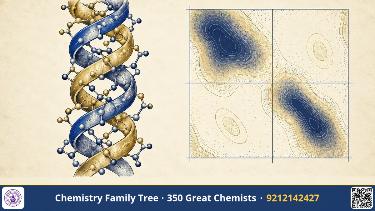
Every biochemistry student meets the Ramachandran plot. It is a simple two-dimensional graph that shows which backbone shapes a protein chain can adopt and which are forbidden by steric clashes. Gopalasamudram Narayanan Ramachandran, working at the University of Madras in the 1950s and 1960s, proposed the triple helical structure of collagen (1954, with Gopinath Kartha) and then introduced the plot that now carries his name (1963, with C. Ramakrishnan and V. Sasisekharan). Both ideas are classic examples of how careful geometry and model building can reveal the shape of biological molecules.
The Scientific Problem Before the Discovery
In 1951 Linus Pauling and Robert Corey proposed the α-helix and β-sheet as the main hydrogen-bonded structures of proteins. Their work depended on a crucial insight: the peptide bond is planar. But there was still a question: how many conformations can a polypeptide chain really adopt? A long chain has many single bonds, so the number of theoretical shapes is enormous. Also, collagen, the most abundant protein in the human body and the main component of skin, tendon and bone, gave X-ray diffraction patterns that did not fit an α-helix. Its structure was a puzzle.
The Discovery: Step by Step
- Step 1: Collagen. Ramachandran and Kartha studied X-ray fibre patterns of collagen and, in 1954, proposed that collagen is made of three polypeptide chains wound together in a triple helix, held together by hydrogen bonds. Glycine is needed at every third position, because only its tiny side chain, a hydrogen atom, fits in the crowded centre.
- Step 2: Refinement. The model was refined by later work, especially by Rich and Crick and by Ramachandran's own group, who corrected details of the hydrogen bonding. The basic idea of a triple helix with a glycine at every third residue remains correct.
- Step 3: Conformational analysis. The planar peptide unit leaves only two rotations per residue: φ (phi) about the N–Cα bond and ψ (psi) about the Cα–C bond. Using hard-sphere models and van der Waals radii, Ramachandran and colleagues calculated which (φ, ψ) pairs avoid atomic overlap.
- Step 4: The plot. They drew the allowed and disallowed regions on a map of φ versus ψ, each from −180° to +180°. This is the Ramachandran map.
Peptide backbone: ...–N–Cα–C(=O)–N–Cα–C(=O)–...
φ (phi): rotation about N–Cα     ψ (psi): rotation about Cα–C
ω (omega): rotation about C–N, about 180° (trans), planar due to partial double bond
The Chemistry Behind It
Planar peptide bond. The lone pair on nitrogen delocalises into the carbonyl group, so the C–N bond has partial double-bond character (about 40 per cent). This makes the six atoms Cα–C(=O)–N(H)–Cα coplanar, usually trans.
Steric hindrance. Many (φ, ψ) combinations bring backbone or side-chain atoms closer than their van der Waals radii allow. These are shown as disallowed regions.
| Structure | Approximate φ, ψ | Notes |
|---|---|---|
| Right-handed α-helix | −57°, −47° | 3.6 residues per turn, i to i+4 hydrogen bonds |
| Antiparallel β-sheet | −139°, +135° | Extended chain |
| Parallel β-sheet | −119°, +113° | Extended chain |
| Left-handed α-helix | about +57°, +47° | Rare, mostly glycine |
| Polyproline II / collagen | about −75°, +145° | Left-handed chains in a right-handed superhelix |
Special residues. Glycine has no side chain so it can adopt a much wider range of φ and ψ. Proline has its side chain bonded to its nitrogen in a ring, which fixes φ near −60°, so it is a helix breaker. This is why a proline-rich, glycine-every-third pattern suits collagen.
Applications Today
- Structure validation. Every protein structure deposited in the Protein Data Bank is checked with a Ramachandran plot. Outliers suggest errors.
- Protein modelling. Homology modelling, molecular dynamics and structure prediction programs use φ and ψ preferences.
- Medicine. Collagen diseases, such as osteogenesis imperfecta, often arise from glycine substitutions that disturb the triple helix. Scurvy results from vitamin C deficiency, which impairs hydroxylation of proline in collagen.
- Materials and cosmetics. Gelatin is partly denatured collagen. Collagen is used in wound dressings and tissue engineering.
- Leather industry. Tanning cross-links collagen. India's Central Leather Research Institute in Chennai worked closely with Ramachandran on collagen.
Exam Corner
- φ is the angle about N–Cα; ψ is the angle about Cα–C; ω is about the peptide bond and is near 180°.
- Glycine allows the widest region of the Ramachandran plot; proline the narrowest.
- Collagen: triple helix of three left-handed polyproline II-type chains coiled into a right-handed superhelix; Gly-X-Y repeats.
- α-helix: 3.6 residues per turn, pitch 5.4 Å, rise 1.5 Å per residue.
- Vitamin C is needed for hydroxylation of proline and lysine in collagen.
Common Misconceptions
- "The Ramachandran plot shows the structure of the protein." It shows backbone dihedral angles of residues and indicates whether they are sterically sensible.
- "The α-helix and collagen helix are the same." They differ greatly. Collagen chains are extended and left-handed, and the three chains twist together into a right-handed supercoil.
- "Forbidden regions are impossible." They are strongly disfavoured, but a few residues, especially glycine, may be found there.
Quick Revision Box
- Collagen triple helix proposed in 1954 by Ramachandran and Kartha.
- Ramachandran plot introduced in 1963 with Ramakrishnan and Sasisekharan.
- φ and ψ are the two free backbone rotations.
- Peptide bond is planar and mostly trans.
- Gly every third residue in collagen; Pro and Hyp are abundant.
- Plot is used for validating protein structures.
FAQ
Did Ramachandran win a Nobel Prize?
No. He was nominated for recognition by many scientists, but did not win a Nobel Prize. His plot is among the most used diagrams in structural biology.
Why is glycine needed in collagen?
Three chains pack so tightly at the centre that only the hydrogen side chain of glycine fits.
What are the axes of the plot?
The horizontal axis is φ and the vertical axis is ψ, both from −180° to +180°.
Is the plot used for all residues?
Separate plots are drawn for glycine, proline and general residues, since their allowed regions differ.
Published by ABC Chemistry — IIT-JAM | CSIR-NET | GATE Chemistry Coaching. Visit abcchemistry.com
About the chemist
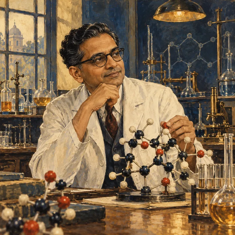
G. N. Ramachandran
1922-2001 · India
Ramachandran plot; collagen triple helix