The discovery, explained · Chemist no. 159 of 350
Hartmut Michel and the First Membrane Protein Crystals: Making the Impossible Possible
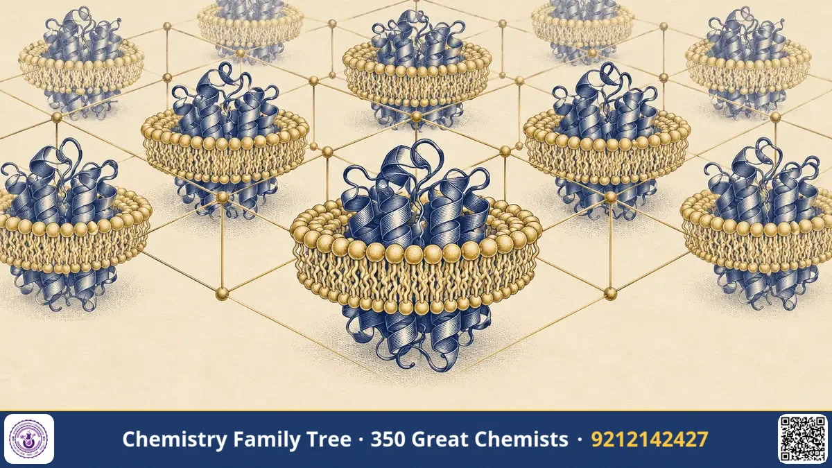
Every cell is wrapped in a membrane, and set into that membrane are proteins that sense signals, pump ions, carry out photosynthesis and burn food with oxygen. These proteins are the targets of a large share of modern medicines. Yet for most of the twentieth century nobody had seen one at atomic resolution, because nobody could grow crystals of them. In 1982, Hartmut Michel grew the first well-ordered three-dimensional crystals of a membrane protein complex, the photosynthetic reaction centre of the purple bacterium Rhodopseudomonas viridis. Together with Johann Deisenhofer and Robert Huber, he was awarded the 1988 Nobel Prize in Chemistry. His story is a lesson in persistence and in the physical chemistry of surfaces.
The Scientific Problem Before the Discovery
X-ray crystallography needs a crystal in which millions of identical molecules sit in the same orientation in a regular lattice. Soluble proteins have a hydrophilic surface and make polar contacts with each other, which is helpful for crystal growth. A membrane protein is different. It has a hydrophobic belt around its middle that interacts with the fatty acid chains of the lipid bilayer, and hydrophilic regions that stick out of the membrane on both sides. If you take the protein out of the membrane and put it in water, the hydrophobic belt is exposed and the proteins aggregate and precipitate.
Before 1980, many researchers believed that membrane proteins would not form three-dimensional crystals at all. The one structural success was bacteriorhodopsin, studied in naturally occurring two-dimensional crystalline patches of the purple membrane by electron microscopy. But a full three-dimensional atomic structure by X-ray diffraction remained out of reach.
The Discovery: Detergents and Careful Chemistry
Michel worked at the Max Planck Institute of Biochemistry in Martinsried, in the group of Dieter Oesterhelt, who studied photosynthetic bacteria and bacteriorhodopsin. Michel decided to try the problem with the reaction centre of Rh. viridis, which is relatively stable. The strategy can be summarised as follows.
- Isolate the protein from the membrane. The bacterial membranes were treated with a detergent. Detergent molecules have a polar head and a hydrophobic tail, and at suitable concentration they form micelles. The tails stick to the hydrophobic belt of the protein, and the heads face the water, so the protein dissolves as a protein–detergent complex.
- Purify the complex. The reaction centre was separated from other proteins and its identity was checked by its absorption spectrum and activity.
- Screen crystallisation conditions. Using vapour diffusion, a drop containing protein, detergent and a precipitant such as ammonium sulfate is allowed to lose water slowly to a larger reservoir. The solution becomes supersaturated and crystals can form.
- Add small amphiphilic molecules. Michel found that adding small amphiphiles such as heptane-1,2,3-triol improved the packing, because these molecules fit in the gaps between large detergent micelles and allow protein surfaces to meet.
- Prove that crystals are of the protein. The crystals, which were deep coloured, were checked to confirm that they contained the intact complex with its pigments.
The crystals diffracted X-rays to high enough resolution for Deisenhofer and Huber to solve the structure. Michel collaborated with them in analysing it and published jointly in the mid-1980s. The structure showed transmembrane helices, with hydrophobic residues facing the lipid and polar groups inside, matching the physical chemistry expected for a membrane protein.
The Chemistry Behind It
Several ideas from your syllabus appear here.
| Concept | Role in membrane protein crystallisation |
|---|---|
| Amphiphiles / surfactants | Detergent molecules with polar head and non-polar tail solubilise membrane proteins |
| Critical micelle concentration | Above this concentration detergent forms micelles |
| Hydrophobic effect | Drives non-polar surfaces together; explains protein aggregation in water |
| Supersaturation | Needed for nucleation and growth of crystals |
| Vapour diffusion | Slow water transfer concentrates the drop |
For a successful crystal, the protein–detergent complex must be homogeneous, and the polar surfaces of the proteins must be able to form contacts. Too large a micelle can block these contacts; this is why a small amphiphile can help.
Michel later worked on another membrane protein, cytochrome c oxidase, the terminal enzyme of respiration. With Iwata and Ostermeier he solved the structure of the enzyme from Paracoccus denitrificans in 1995 using a trick: an antibody fragment bound to the protein provided extra polar surface for crystal contacts. The enzyme catalyses the reduction of oxygen to water, coupled with proton pumping:
O₂ + 4 cyt c(Fe²⁺) + 4 H⁺ → 2 H₂O + 4 cyt c(Fe³⁺)
Applications Today
- Drug discovery: Many drugs act on membrane proteins such as G protein-coupled receptors, ion channels and transporters. Structures of these targets use crystallisation strategies descended from Michel's work.
- Antibiotic and antiviral research: Efflux pumps and viral membrane proteins are studied structurally.
- Bioenergetics: Structures of respiratory and photosynthetic complexes explain how electron transfer is coupled with proton pumping.
- Biotechnology: Detergents and lipid-like media (such as lipidic cubic phases, developed by other scientists) are standard in membrane protein labs.
- India: Structural biology groups in IISc, the IISERs and other institutes work on membrane protein targets relevant to tuberculosis and malaria.
Exam Corner
- Hartmut Michel shared the 1988 Nobel Prize in Chemistry with Deisenhofer and Huber.
- He crystallised the photosynthetic reaction centre of Rhodopseudomonas viridis in 1982.
- Detergents are amphiphilic: polar head, hydrophobic tail; they form micelles above the CMC.
- Cytochrome c oxidase: O₂ + 4e⁻ + 4H⁺ → 2H₂O with proton pumping across the membrane.
- Transmembrane segments are often α-helices with hydrophobic outer surfaces.
Common Misconceptions
- "Detergent destroys the protein." Mild detergents keep membrane proteins intact by replacing the lipids around them.
- "Michel solved the structure by himself." He made the crystals and joined the analysis; the structure solution was done with Deisenhofer and Huber.
- "A micelle is the same as a bilayer." A micelle is a small sphere or disc of detergent; a bilayer is a flat double layer of lipids.
Quick Revision Box
- Problem: membrane proteins have a hydrophobic belt, aggregate in water.
- Solution: mild detergent micelles keep them soluble; small amphiphiles improve packing.
- 1982: first 3D crystals of a membrane protein complex (reaction centre).
- 1995: cytochrome c oxidase structure from Paracoccus denitrificans.
- Cyt c oxidase: O₂ + 4e⁻ + 4H⁺ → 2H₂O.
- 1988 Nobel Prize in Chemistry.
FAQ
Why are membrane proteins difficult to crystallise?
Their hydrophobic surfaces make them aggregate in water, and detergent micelles around them can hinder regular packing.
What is a detergent micelle?
A small aggregate of detergent molecules with hydrophobic tails inside and polar heads outside, which can surround a membrane protein.
Did Michel work with Deisenhofer and Huber?
Yes. Michel provided the crystals and took part in the structure analysis at Martinsried.
Is this topic in the exam syllabus?
Surfactants, micelles and the hydrophobic effect are part of physical chemistry, and enzyme and X-ray topics appear in biochemistry sections.
Published by ABC Chemistry — IIT-JAM | CSIR-NET | GATE Chemistry Coaching. Visit abcchemistry.com
About the chemist
Hartmut Michel
1948- · Germany · Nobel Chemistry 1988
Crystallized photosynthetic reaction centre