The discovery, explained · Chemist no. 135 of 350
John Kendrew and Myoglobin: The First Three-Dimensional Picture of a Protein
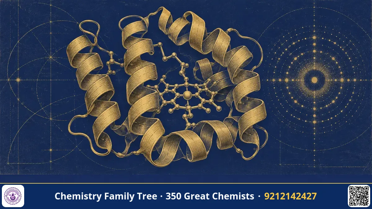
Until 1957, nobody had ever seen what a protein really looks like. Chemists knew proteins as long chains of amino acids, but how those chains fold in space was a mystery. At the Cavendish Laboratory in Cambridge, John Cowdery Kendrew and his colleagues solved this problem for the oxygen-storing protein myoglobin from the sperm whale. Using X-ray crystallography and years of patient computation, he produced the first atomic-level model of a protein. The result showed an unexpectedly irregular, compact fold, packed with α-helices around an iron-containing heme group. In 1962 Kendrew shared the Nobel Prize in Chemistry with Max Perutz "for their studies of the structures of globular proteins". This post explains the method and the biochemical meaning.
The Scientific Problem Before the Discovery
Linus Pauling had proposed the α-helix and β-sheet in 1951 as likely structural elements of proteins, based on hydrogen bonding in the peptide backbone. But these were models, not observations, and no one knew how helices combine into a working protein. X-ray diffraction had solved the structures of small molecules such as salts and simple organic compounds, but a protein has thousands of atoms. The diffraction pattern of a protein crystal contains thousands of spots, and each spot has an intensity (easy to measure) and a phase (impossible to measure directly). Without the phases, you cannot compute the electron density map. This is the famous phase problem.
The Discovery: Step by Step
- Choosing the protein. Kendrew chose myoglobin because it is small (about 153 amino acids and a molecular mass near 17,000), and because diving mammals such as the sperm whale have a lot of it in their muscles, which allows a plentiful supply. It crystallised well.
- Growing crystals and collecting data. Crystals were X-rayed, and the positions and intensities of diffraction spots recorded on film. Kendrew's team collected thousands of reflections.
- Solving the phase problem. Using the method of isomorphous replacement, which Max Perutz had developed for haemoglobin, heavy atoms such as mercury or gold were attached to specific sites on the protein without changing its fold. The difference in the diffraction intensities between native and heavy-atom crystals gave the phases.
- Low-resolution map (1957). A 6 Å electron density map showed a compact molecule with rod-like segments (helices) and a flat disc of heme. This was the first view of a protein structure, described as a "sausage-like" tangle, to the surprise of many who expected a regular, symmetrical object.
- High-resolution map (1959-60). With the use of a computer (the Cambridge EDSAC machine), the resolution was improved to 2 Å, where individual amino acid side chains and the path of the polypeptide chain could be traced.
The key mathematical tool is the Fourier transform, which converts diffraction data into the electron density:
ρ(x,y,z) = (1/V) Σ F(h,k,l) · exp[−2πi(hx + ky + lz)]
Here F(h,k,l) is the structure factor, including both amplitude and phase. The geometry of diffraction is governed by Bragg's law, nλ = 2d sin θ.
The Chemistry Behind It
Myoglobin consists of a single polypeptide chain folded into eight α-helical segments (labelled A to H) joined by short non-helical turns. About three quarters of the chain is helical. Key findings:
- Hydrophobic core. Non-polar side chains (Leu, Val, Phe) are inside; polar and charged side chains are on the outside, facing water. This showed that hydrophobic interactions drive protein folding.
- Heme pocket. The iron(II) atom of the heme sits in a pocket. It is bonded to four nitrogen atoms of the porphyrin ring and to the imidazole nitrogen of a histidine (the proximal histidine, His F8). The sixth position binds oxygen reversibly. A second histidine (distal, His E7) sits nearby.
- Reversible oxygen binding:
Mb + O₂ ⇌ MbO₂. The oxygen-binding curve of myoglobin is a simple hyperbola, with high oxygen affinity, so it stores oxygen in muscle. Haemoglobin, a tetramer, shows a sigmoid curve because of cooperativity. - Primary structure decides the fold. The structure confirmed that the amino acid sequence contains the information for three-dimensional shape, in line with Anfinsen's later experiments.
In the same period Perutz solved the structure of haemoglobin, and comparing the two showed that the globin fold is conserved, evidence of a common evolutionary origin.
Applications Today
- Structure-based drug design. Modern drugs such as HIV protease inhibitors and kinase inhibitors for cancer were designed after solving protein structures by crystallography.
- Protein Data Bank. Today more than 200,000 structures are in the PDB, and myoglobin was among the earliest entries.
- Molecular biology. Understanding enzymes, receptors and antibodies depends on crystal structures; cryo-electron microscopy and AlphaFold prediction add to this tradition.
- Medicine. Sickle cell disease is understood as a one-amino-acid change in haemoglobin; structural biology explains how such mutations alter protein shape.
- India. Indian scientist G. N. Ramachandran introduced the Ramachandran plot (1963), which is still used to check the backbone geometry of protein structures, and protein crystallography is practised at leading Indian institutes.
Exam Corner
- Kendrew solved the structure of sperm whale myoglobin (1957-60); Perutz solved haemoglobin; 1962 Nobel Prize in Chemistry.
- Myoglobin: single chain of 153 amino acids, eight α-helices, one heme, one O₂ binding site.
- Fe²⁺ binds His F8 (proximal) and O₂; hyperbolic oxygen binding curve.
- Phase problem solved by isomorphous replacement using heavy atoms.
- Bragg's law: nλ = 2d sin θ.
Common Misconceptions
- "Kendrew discovered the α-helix." That was Pauling, Corey and Branson (1951). Kendrew's structure was the first experimental observation of helices in a real protein.
- "Myoglobin and haemoglobin are the same." Myoglobin is a monomer for oxygen storage; haemoglobin is a tetramer for oxygen transport, showing cooperativity.
- "Kendrew won a Nobel Prize in Medicine." His prize was in Chemistry.
Quick Revision Box
- First protein structure: sperm whale myoglobin, John Kendrew, 1957-1960.
- Method: X-ray diffraction, Fourier synthesis, isomorphous replacement.
- Eight α-helices (A-H), heme with Fe²⁺, 153 residues.
- Hydrophobic residues inside, polar residues outside.
- Haemoglobin structure was solved by Max Perutz.
- Nobel Prize in Chemistry 1962, shared with Perutz.
FAQ
Why was myoglobin chosen?
It is small, easily crystallised and abundant in sperm whale muscle.
What is the phase problem?
X-ray detectors record only the intensity of diffracted beams, not their phases, which are required to calculate the electron density map.
How does myoglobin bind oxygen?
Oxygen binds reversibly to the Fe²⁺ of the heme group in a protein pocket.
Who shared the Nobel Prize with Kendrew?
Max Perutz, for the structure of haemoglobin.
Published by ABC Chemistry — IIT-JAM | CSIR-NET | GATE Chemistry Coaching. Visit abcchemistry.com
About the chemist
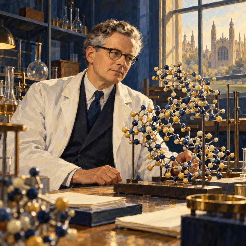
John Kendrew
1917-1997 · United Kingdom · Nobel Chemistry 1962
Structure of myoglobin