The discovery, explained · Chemist no. 189 of 350
Martin Chalfie and the Glowing Worm: GFP Becomes a Gene Marker
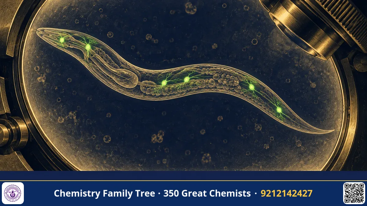
In 1994 a cover image of a tiny transparent roundworm, with a few neurons glowing bright green, announced a new era in biology. The picture came from the laboratory of Martin Chalfie at Columbia University, who showed that the green fluorescent protein (GFP) of the jellyfish Aequorea victoria, discovered by Osamu Shimomura, works as a genetic marker in other organisms. By attaching the GFP gene to the control region of another gene, biologists could see in a living animal exactly where and when that gene is switched on.
For this, Chalfie shared the 2008 Nobel Prize in Chemistry with Shimomura and Roger Tsien. Chalfie himself is a neurobiologist, but the prize recognises a chemical tool: a protein whose fluorescence needs no additional cofactor.
The Scientific Problem Before the Discovery
By 1990 molecular biologists could clone genes and tell the sequence of DNA, but seeing a gene's activity inside a living organism was hard. Existing reporter genes had limits:
- β-Galactosidase (lacZ): The enzyme converts the substrate X-gal into a blue dye, but the cells must be killed and fixed, and the substrate must be supplied.
- Luciferase: Needs the substrate luciferin and ATP and gives only faint light.
- Antibodies: They show where a protein is but require fixation of dead tissue.
Chalfie studied the nematode worm Caenorhabditis elegans, an animal about 1 mm long with only 302 neurons. It is transparent, so one can look at living cells. He studied the six touch-receptor neurons that let the worm feel a gentle touch, and he wanted to watch these cells in living animals. He needed a marker that would be visible with no added chemicals.
The Discovery: Step by Step
- The idea (1989): Chalfie attended a seminar on bioluminescence and heard about GFP, a protein that fluoresces green under blue or UV light. He realised that if it made its chromophore without special enzymes, it could be a universal marker. Nobody knew whether it would work in other species.
- Getting the gene: Douglas Prasher, a molecular biologist at Woods Hole, had cloned a cDNA of GFP and shared it with Chalfie's laboratory. Chalfie's group needed only the coding sequence.
- Test in bacteria: A student, Ghia Euskirchen, inserted the gene (amplified by PCR) into E. coli under an inducible promoter. The cells glowed green under long-wave UV light, even though bacteria had no jellyfish enzymes.
- Test in worms: The GFP gene was fused to the promoter of the mec-7 gene, which is switched on in the touch-receptor neurons. The construct was injected into worms, and six touch cells appeared as green cells, including their long processes.
- Publication (1994): The results were published in the journal Science as "Green fluorescent protein as a marker for gene expression", and requests for the plasmid soon flooded in from other laboratories.
The Chemistry Behind It
The key chemical fact: GFP's chromophore is formed autocatalytically inside the protein once it folds, with only O₂ from air as a co-reactant. The three residues Ser65, Tyr66 and Gly67 cyclise, the backbone nitrogen of Gly67 attacks the carbonyl of Ser65, followed by dehydration and oxidation (loss of two hydrogens) of the Tyr66 side chain, producing a conjugated 4-(p-hydroxybenzylidene)imidazolin-5-one:
Ser65–Tyr66–Gly67 →(fold, cyclise, –H₂O) imidazolinone →(O₂, –H₂O₂) conjugated chromophore
Absorption occurs at about 395 nm (neutral form) and 475 nm (anionic form), and green emission peaks near 509 nm. The difference between absorption and emission wavelengths is the Stokes shift, caused by relaxation of the excited state and rearrangement of hydrogen bonding in the protein. The β-barrel shields the chromophore from collisions with water that would quench fluorescence, which gives a high quantum yield.
The reporter gene concept can be written as a construct:
[promoter of gene X] — [GFP coding sequence] — [terminator]
GFP appears only in cells where gene X's promoter is active.
Also, GFP can be fused to the coding sequence of a protein itself (Protein–GFP fusion), so that one sees where the protein is located within the cell, for example in the nucleus or mitochondria. Because the barrel has its N and C termini close to each other on the surface, such fusions usually keep both protein and fluorescence.
Applications Today
- Neuroscience: Mapping neurons and circuits in worms, flies, fish and mice.
- Cancer research: Tracking metastasis by labelling tumour cells with GFP.
- Developmental biology: Watching which cells express a gene at each stage of the embryo.
- Infectious disease: GFP-tagged bacteria, malaria parasites and viruses can be followed in living hosts. India's research institutes use these tools to study tuberculosis and malaria.
- Biotechnology: Screening of transformed cells and plants, e.g., in genetic engineering practicals.
Exam Corner
- Chalfie shared the 2008 Nobel Prize in Chemistry with Shimomura and Tsien for the discovery and development of GFP.
- Chalfie showed GFP fluoresces in E. coli and in C. elegans touch neurons (1994).
- A reporter gene is fused to a promoter to show where and when the gene is active.
- GFP chromophore forms spontaneously from Ser65-Tyr66-Gly67 with O₂; no cofactors.
- C. elegans has 302 neurons and is transparent, ideal for imaging.
Common Misconceptions
- "Chalfie discovered GFP." Shimomura discovered GFP; Prasher cloned the gene; Chalfie showed it could be used as a marker in other organisms.
- "GFP needs a substrate to glow." It needs only light and, for forming its chromophore, oxygen.
- "Worms glow in the dark." They fluoresce only when excited by blue or UV light.
Quick Revision Box
- Martin Chalfie, Columbia University; Nobel Prize in Chemistry 2008 (shared).
- Showed GFP works in bacteria and the worm C. elegans (1994).
- Gene cloned earlier by Douglas Prasher.
- Used the mec-7 promoter to label touch receptor neurons.
- GFP: ex ~395/475 nm, em ~509 nm; barrel of 11 strands.
- Concept: reporter gene or fusion tag.
FAQ
Why is C. elegans a good model?
It is transparent, small, easy to grow, and its entire cell lineage and nervous system are mapped.
What is a promoter?
A DNA region where RNA polymerase begins transcription; it controls where and when a gene is active.
Why did the experiment surprise people?
Many thought GFP would need other jellyfish-specific enzymes to form its chromophore, but it worked in bacteria and worms.
What is a protein fusion?
A gene engineered so that two proteins are made as a single chain, such as a protein of interest joined to GFP, so its location can be seen.
Published by ABC Chemistry — IIT-JAM | CSIR-NET | GATE Chemistry Coaching. Visit abcchemistry.com

About the chemist
Martin Chalfie
1947- · United States · Nobel Chemistry 2008
GFP as a biological marker