The discovery, explained · Chemist no. 323 of 350
Maurice Wilkins and the X-ray Evidence for the DNA Helix
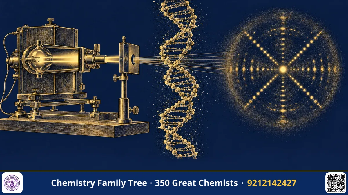
Before anyone could build a model of DNA, someone had to show what the real molecule looked like. At King's College London, Maurice Wilkins (1916-2004) led a group that obtained X-ray diffraction patterns of DNA fibres which strongly suggested that the molecule is a regular helix. Working with Raymond Gosling and, from 1951, Rosalind Franklin, he helped produce the pictures and measurements that Watson and Crick used to build their double-helix model in 1953. For his role, Wilkins shared the 1962 Nobel Prize in Physiology or Medicine with Watson and Crick. This blog explains in simple terms how X-ray diffraction reveals molecular shape, and what the DNA patterns told us.
The Scientific Problem Before the Discovery
By the late 1940s it was believed that DNA carried genetic information, but its structure was unknown. The chemical building blocks, nucleotides linked by phosphodiester bonds, had been worked out by Alexander Todd and others. The sizes of DNA molecules were known to be very large. But the way these long chains are arranged in space was a mystery, and without this nobody could understand how genes copy themselves.
Wilkins, trained as a physicist, had worked on the Manhattan Project in the United States on isotope separation. After the war, shocked by the use of atomic weapons, he turned to biophysics. In Edinburgh and then at King's College London in the Medical Research Council's Biophysics Unit under John Randall, he began to study DNA by physical methods, such as ultraviolet microscopy and, later, X-ray diffraction.
The Discovery / Experiment
Step 1: Getting DNA fibres
In 1950 Wilkins obtained a very pure, highly concentrated gel of DNA from the Swiss chemist Rudolf Signer. Touching a glass rod to the gel and pulling it away drew out thin, uniform fibres of DNA, all with their long molecules lying parallel to the fibre axis. Wilkins and Gosling noticed that these fibres were so regular and clear, under a polarising microscope, that they suggested a well-ordered structure inside.
Step 2: Principle of X-ray diffraction
When a beam of X-rays strikes a regular arrangement of atoms, the rays are scattered and interfere. Where the waves are in phase, spots or streaks appear on a photographic film. The condition for reinforcement is given by Bragg's law:
nλ = 2d sin θ
(n = order, λ = wavelength, d = spacing between planes, θ = angle)
Larger spacings in the object give diffraction at smaller angles, so the pattern is like a reciprocal image of the structure. For a helix, the pattern has a characteristic cross shape (X) with horizontal layer lines. Cochran, Crick and Vand published the mathematical theory of helical diffraction in 1952, showing how the positions of the layer lines relate to the pitch of the helix and the cross shape to its radius.
Step 3: The first DNA pictures
In 1950-51 Wilkins and Gosling took X-ray pictures of DNA fibres with fine beams. At a meeting in Naples in May 1951, Wilkins showed a pattern that appeared crystalline, and James Watson, who was present, became inspired to work on DNA. Wilkins's group found that the DNA fibres gave a sharp, crystalline pattern in dry air (the A form) and a more diffuse helical pattern when kept moist (the B form).
Step 4: Franklin's improvements and Photo 51
Rosalind Franklin, an expert in X-ray crystallography, joined King's in January 1951, with Gosling as her student. She refined the technique by controlling the humidity precisely, and by using a fine-focus X-ray tube and a microcamera. She separated the A and B forms clearly. In May 1952 Franklin and Gosling took the sharp B-form picture known as Photo 51, with a clear X pattern. In January 1953 Wilkins showed this photograph to Watson. Together with Franklin's unpublished report to the Medical Research Council, passed on to Crick by Max Perutz, it provided the key data, but without Franklin's permission. The relationship between Wilkins and Franklin was strained, partly because of a misunderstanding about her role in the group at the start.
Step 5: What the pattern showed
- The cross shape: DNA is helical.
- Layer lines spaced for a repeat distance of 34 Å: one full turn of the helix.
- A strong reflection at 3.4 Å: base pairs stacked at that distance, so about 10 base pairs per turn.
- The width of about 20 Å: two chains, with the phosphates on the outside.
- Missing fourth layer line: pointed to two strands offset from each other, which Crick interpreted as a double helix.
The Chemistry Behind It
- Water content and forms: At relative humidity of about 75 percent or less, DNA takes the A form (11 base pairs per turn, tilted bases). At high humidity it takes the B form (10 base pairs per turn, bases perpendicular to the axis), which is the form inside cells.
- Sodium salt: Wilkins's DNA was the sodium salt, with Na+ ions neutralising the negative phosphate charges.
- Fibre diffraction: Fibres are not perfect crystals; the diffraction shows rotational averaging, and thus gives less detail than single-crystal diffraction, but enough for the helix parameters.
- Later confirmation: Wilkins and colleagues, including Stokes and Wilson, confirmed the Watson-Crick model by more X-ray work in 1953, and extended it to RNA and nucleoproteins.
Applications Today
- Structural biology: X-ray crystallography is used to determine structures of proteins, DNA and drug targets, including at Indian centres such as IISc Bangalore and the synchrotron facility at RRCAT Indore.
- Drug design: Structures of enzymes and receptors bound to inhibitors guide drug design, e.g. HIV protease inhibitors.
- Materials science: Powder XRD identifies phases of solids, catalysts and nanomaterials, a core topic in M.Sc. chemistry.
- Forensic and pharmaceutical analysis: XRD fingerprints crystalline drugs and polymorphs.
- Complementary methods: NMR and cryo-electron microscopy now complement X-rays for large complexes.
Exam Corner
- Bragg's equation: nλ = 2d sinθ.
- Wilkins, Watson and Crick shared the 1962 Nobel Prize in Physiology or Medicine.
- B-DNA: pitch 34 Å, rise 3.4 Å, diameter 20 Å, 10 bp per turn; A-DNA: 11 bp per turn.
- Photo 51 was taken by Franklin and Gosling in 1952 and showed the X-shaped pattern of a helix.
- Helical diffraction theory was given by Cochran, Crick and Vand (1952).
Common Misconceptions
- "Wilkins took Photo 51." It was taken by Franklin and her student Gosling. Wilkins showed it to Watson.
- "X-ray diffraction gives a direct photograph of the molecule." It gives a diffraction pattern, which has to be interpreted mathematically to deduce the structure.
- "Wilkins won the Chemistry Nobel." His prize was in Physiology or Medicine, and he was a physicist by training.
Quick Revision Box
- DNA fibres from Signer's gel: parallel molecules, ideal for diffraction.
- Bragg's law: nλ = 2d sinθ.
- X pattern = helix; 34 Å pitch; 3.4 Å base spacing.
- A form (dry) and B form (wet) found at King's College.
- Photo 51 (1952): Franklin and Gosling.
- Nobel Prize 1962, Physiology or Medicine.
FAQ
What role did Wilkins play in finding the DNA structure?
He led the King's College X-ray work on DNA fibres, obtained early diffraction pictures, shared data with Watson and Crick and later confirmed the model.
Why is a fibre good enough for DNA diffraction?
Long DNA molecules align along the fibre axis, so the pattern shows helical repeats even though the sample is not a single crystal.
Why is Rosalind Franklin not on the Nobel Prize?
She died in 1958 and the Nobel Prize is not awarded after death. In any case, the 1962 prize was limited to three laureates.
Is this topic useful for chemistry exams?
Yes. Bragg's law, diffraction, and the dimensions of A, B and Z-DNA are common in JAM, NET and GATE questions.
Published by ABC Chemistry — IIT-JAM | CSIR-NET | GATE Chemistry Coaching. Visit abcchemistry.com
About the chemist
Maurice Wilkins
1916-2004 · United Kingdom · Nobel Physiology/Medicine 1962
X-ray diffraction studies of DNA