The discovery, explained · Chemist no. 68 of 350
Max Perutz and the Structure of Haemoglobin: How Blood Carries Oxygen
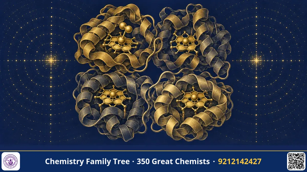
Every breath you take ends with a chemical reaction in your red blood cells. Oxygen sticks to haemoglobin in the lungs and is released in the tissues, and the way this happens is neither random nor simple. Max Perutz spent about 22 years solving the three-dimensional structure of haemoglobin, a protein of roughly 64,500 daltons, and then used the structure to explain how it works. He shared the 1962 Nobel Prize in Chemistry with John Kendrew. His work showed that a protein is a precisely folded machine, and that small changes at one site can change the behaviour of distant sites. This idea is called allostery, and it remains central in biochemistry.
The Scientific Problem Before the Discovery
By the 1930s haemoglobin was one of the best-studied proteins. Scientists knew that it contained iron, that it carried oxygen, and that its oxygen-binding curve was S-shaped (sigmoidal). Muscle myoglobin, a related protein, showed a simple hyperbolic curve. The question was why haemoglobin behaved differently. Chemists suspected that four oxygen-binding sites interacted with each other, but without a structure no one could say how.
The size of the problem was daunting. Haemoglobin has nearly 600 amino acid residues and thousands of atoms. Using X-rays on such a crystal gives thousands of spots, and in the 1930s there were no computers to process them. Many physicists and chemists thought the task was impossible.
The Discovery: Step-by-Step
- Crystals and first photographs (1937). Perutz began work at Cambridge in the Cavendish Laboratory under J. D. Bernal and W. L. Bragg, using horse haemoglobin crystals.
- The phase problem. Detectors record intensities of diffracted X-rays but not phases, and without phases one cannot calculate the electron density map.
- Isomorphous replacement (1953). Perutz found that heavy atoms such as mercury could be attached to specific sites in the protein without changing the crystal's shape. Comparing the diffraction pattern before and after attaching the heavy atoms gave the phase information.
- Low-resolution map (1959). The first three-dimensional model at 5.5 Å resolution showed four subunits arranged in a compact tetramer.
- High-resolution map (1960s). Improved data at about 2.8 Å allowed him to fit amino acid side chains and see how the iron in each heme group was held.
Meanwhile, Perutz's colleague John Kendrew had solved the structure of the simpler protein myoglobin in 1958 at 6 Å, then at higher resolution. Seeing that the chains of haemoglobin folded like myoglobin gave early confirmation that the work was correct.
The Chemistry Behind It
Haemoglobin is a tetramer with two α chains and two β chains (α2β2). Each chain carries one heme, which is an iron(II) ion held by four nitrogen atoms of a porphyrin ring. A histidine side chain from the protein (the proximal histidine) binds the iron on one side, and oxygen binds on the opposite side:
Hb + 4O2 ⇌ Hb(O2)4
In the deoxy form, high-spin Fe(II) is slightly too large to fit into the porphyrin plane, so it sits about 0.4 Å out of the plane. When oxygen binds, iron becomes effectively low-spin and smaller, and it moves into the plane. This movement pulls on the proximal histidine and changes the arrangement of the helix, which shifts the contacts between subunits. Perutz described two quaternary states:
- T state (tense): low oxygen affinity, stabilised by salt bridges between subunits.
- R state (relaxed): high oxygen affinity, where some salt bridges are broken.
Binding of the first oxygen makes it easier for the next to bind. This is positive cooperativity, giving the sigmoidal curve. The Hill equation describes it: with Hill coefficient n, fractional saturation is Y = [O2]^n / (P50^n + [O2]^n). For haemoglobin n is about 2.8, while for myoglobin n = 1.
Other effects can also be explained. In the Bohr effect, lower pH and higher CO2 reduce oxygen affinity so that tissues get more oxygen. The molecule 2,3-bisphosphoglycerate (2,3-BPG) binds in the central cavity between the β chains of the T state and also lowers oxygen affinity. Perutz also used the structure to explain sickle cell anaemia: the mutation replaces glutamic acid with valine at position 6 of the β chain, creating a sticky hydrophobic patch that causes the haemoglobin to form fibres.
Applications Today
- Understanding disease: Sickle cell anaemia and thalassaemia, which are common in parts of India, are better understood from the structure.
- Blood substitutes: Research on artificial oxygen carriers builds on the principles of haemoglobin function.
- Drug design: Allosteric drugs, which bind away from the active site, follow the logic that Perutz demonstrated.
- Medicine and physiology: Altitude adaptation, carbon monoxide poisoning and pulse oximetry can all be explained by oxygen-binding chemistry.
Exam Corner
- Haemoglobin is an α2β2 tetramer containing four heme groups, each with one Fe(II).
- Oxygen binding is cooperative; the curve is sigmoidal, unlike the hyperbolic curve of myoglobin.
- Hill coefficient of haemoglobin is about 2.8 (less than 4, the number of binding sites).
- The Bohr effect: lower pH or more CO2 shifts the curve to the right (lower affinity).
- Perutz shared the 1962 Chemistry Nobel with John Kendrew; Kendrew solved myoglobin.
Common Misconceptions
- "Each oxygen binds independently." No. Binding sites communicate through subunit interactions.
- "Carbon monoxide binds to a different site." CO binds to the same iron, but about 200 times more strongly than oxygen, which is why it is so dangerous.
- "Iron changes from Fe(II) to Fe(III) when it carries oxygen." In normal oxygen transport the iron stays Fe(II). Oxidation to Fe(III) gives methaemoglobin, which cannot carry oxygen.
Quick Revision Box
- Haemoglobin: α2β2 tetramer, four heme-Fe(II) sites.
- T state = low affinity; R state = high affinity.
- Fe moves into the porphyrin plane on oxygenation.
- Heavy-atom isomorphous replacement solved the phase problem.
- Bohr effect and 2,3-BPG lower oxygen affinity.
- Nobel Prize in Chemistry 1962 (with Kendrew).
FAQ
What is cooperativity?
It is when binding of a ligand at one site changes the affinity of other sites on the same molecule. In haemoglobin, binding of the first oxygen raises the affinity for the next.
Why did the structure take so long?
The molecule is large, the data are huge, and the phase problem had to be solved first. Computers capable of the Fourier calculations were not available for years.
What is the difference between myoglobin and haemoglobin?
Myoglobin is a single-chain protein that stores oxygen in muscle. Haemoglobin is a tetramer in blood that transports oxygen with cooperative behaviour.
What is the proximal histidine?
It is the histidine residue that coordinates directly to the iron on the side opposite to oxygen. Its movement transmits the signal between subunits.
Published by ABC Chemistry — IIT-JAM | CSIR-NET | GATE Chemistry Coaching. Visit abcchemistry.com
About the chemist
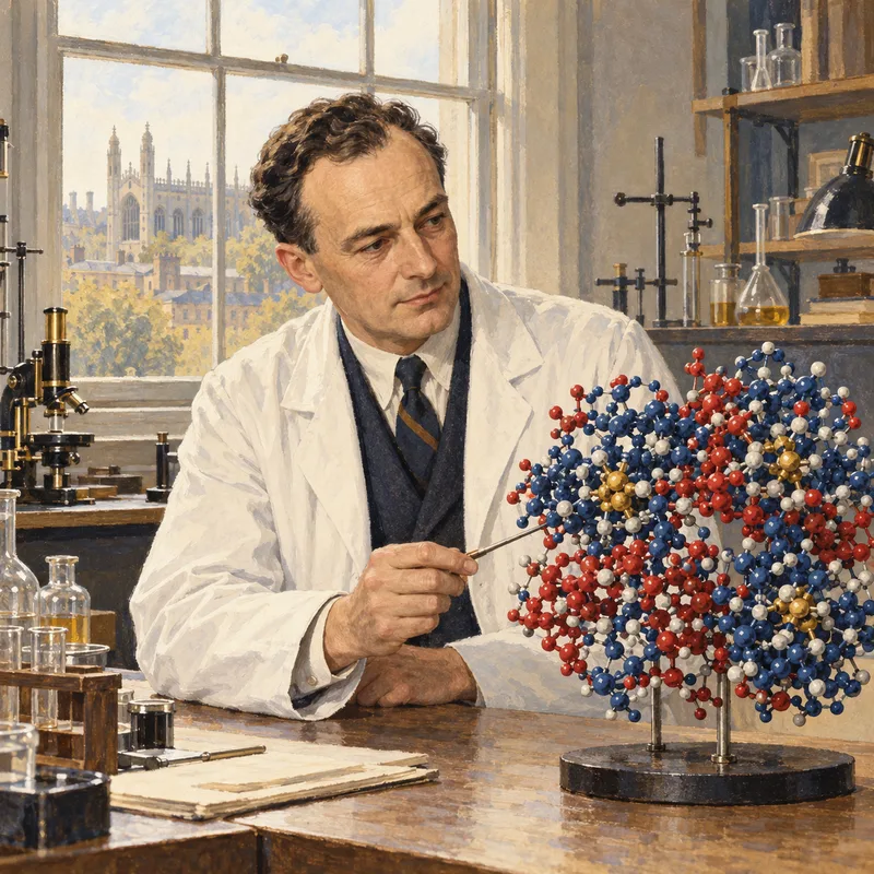
Max Perutz
1914-2002 · Austria/United Kingdom · Nobel Chemistry 1962
Structure of haemoglobin