The discovery, explained · Chemist no. 158 of 350
Robert Huber and Protein Crystallography: From Trypsin Inhibitor to the Reaction Centre
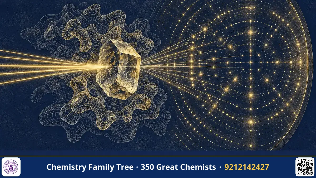
Proteins do almost everything in a cell, but they are far too small to be seen even with the best light microscope. Robert Huber spent a career turning X-ray diffraction patterns into atomic pictures of these molecules. In his laboratory at the Max Planck Institute of Biochemistry in Martinsried, near Munich, he and his colleagues solved the structures of enzymes, antibodies, inhibitors and, together with Johann Deisenhofer and Hartmut Michel, the photosynthetic reaction centre. This earned them the 1988 Nobel Prize in Chemistry. Huber's contributions show how three-dimensional structure explains function, a principle behind modern drug design.
The Scientific Problem Before the Discovery
In the 1950s and 1960s Max Perutz and John Kendrew solved the first protein structures, haemoglobin and myoglobin, after many years of effort. The path was slow because of the phase problem: the diffraction experiment measures the intensity of reflections but not their phases. For proteins, the standard solution was multiple isomorphous replacement, in which heavy atoms such as mercury or platinum are soaked into the crystal. Preparing such derivatives, collecting thousands of reflections and building a model by hand were laborious.
There was also a biological challenge. Chemists wanted to understand how enzymes recognise substrates, how inhibitors block enzymes, how antibodies bind antigens and how pigments in photosynthesis capture light. Each of these needed a detailed structure, preferably at a resolution where side chains and bound ligands could be seen.
The Work: From Crystals to Atomic Models
Huber's group developed and refined many techniques of protein crystallography and applied them to important problems. The typical pipeline in his laboratory included:
- Purification and crystallisation. The protein is purified and slowly concentrated under conditions that encourage ordered crystals.
- X-ray diffraction. A crystal is exposed to X-rays, and the diffraction spots are recorded. Bragg's law, nλ = 2d sinθ, relates the spot positions to the unit cell.
- Phasing. Phases are obtained using heavy-atom derivatives or other methods.
- Electron density map. A Fourier transform of amplitudes and phases gives ρ(x, y, z), a three-dimensional map.
- Model building and refinement. The amino acid sequence is fitted to the map, and the model is improved to agree better with the data.
Important structures from his group include:
- Bovine pancreatic trypsin inhibitor (BPTI) and its complex with trypsin. This showed how a small protein blocks a serine protease by presenting a loop that mimics a substrate.
- Immunoglobulin fragments: Early structures of antibody parts showing the domain fold of antibodies.
- The reaction centre of Rhodopseudomonas viridis: solved with Deisenhofer, using crystals obtained by Michel, the first membrane protein complex structure.
- Later work on proteolytic enzymes and on the proteasome, the large barrel-shaped complex that degrades proteins in the cell.
The Chemistry Behind It
The serine protease trypsin cuts peptide bonds after positively charged residues, lysine or arginine. Its active site contains the catalytic triad Ser195, His57 and Asp102. In simplified form, His57 acts as a base to remove a proton from Ser195, and the serine oxygen attacks the carbonyl carbon of the peptide bond, forming a tetrahedral intermediate:
Ser–OH + R–CO–NHR′ → Ser–O–C(O⁻)(R)–NH₂⁺R′ (tetrahedral intermediate) → acyl-enzyme + R′NH₂
Trypsin inhibitors bind in the active site in a substrate-like manner but are cleaved extremely slowly, so they act as tight-binding inhibitors. Structures like those solved in Huber's group helped define this picture of enzyme–inhibitor recognition.
| Technique element | Purpose |
|---|---|
| Bragg's law | Relates diffraction angle to lattice spacing |
| Heavy-atom derivative | Provides phase information |
| Fourier transform | Converts structure factors to electron density |
| Refinement | Improves the fit between model and data |
Applications Today
- Drug design: Structure-based design of protease inhibitors, including drugs for blood pressure and viral infections, relies on structures of enzymes with bound inhibitors.
- Cancer therapy: Proteasome inhibitors are used to treat multiple myeloma; the structure of the proteasome was important to understanding them.
- Antibody engineering: Knowledge of antibody domains is used in making therapeutic antibodies.
- Energy research: The reaction centre structure inspires work on artificial photosynthesis.
- Academic training: Indian laboratories use similar crystallographic pipelines in structural biology, and the Protein Data Bank is open to all students.
Exam Corner
- Robert Huber shared the 1988 Nobel Prize in Chemistry with Johann Deisenhofer and Hartmut Michel.
- Serine protease catalytic triad: Ser, His, Asp.
- Protein crystallography uses Bragg's law and Fourier synthesis; phases come from isomorphous replacement or other methods.
- BPTI is a small protein inhibitor of trypsin.
- Proteasome: multi-subunit protease complex that degrades proteins tagged with ubiquitin.
Common Misconceptions
- "Crystal structures show proteins as rigid." Crystals give an averaged snapshot; proteins move in solution, and other methods study dynamics.
- "All protein crystals are easy to grow." Crystallisation is often the hardest step.
- "Huber won for the proteasome." The 1988 prize was for the reaction centre; the proteasome came later in his career.
Quick Revision Box
- Huber: Max Planck Institute of Biochemistry, Martinsried; protein crystallographer.
- Solved trypsin–BPTI complex, antibody fragments, reaction centre, proteasome.
- Crystallography steps: crystallise, diffract, phase, map, build, refine.
- Catalytic triad of trypsin: Ser195, His57, Asp102.
- 1988 Nobel Prize in Chemistry, shared with Deisenhofer and Michel.
- Structure explains function: key to drug design.
FAQ
What is the Protein Data Bank?
It is a free archive of three-dimensional structures of proteins and nucleic acids, started in the 1970s, to which structures from Huber's group were also deposited.
How does a trypsin inhibitor work?
It binds the active site like a substrate but the peptide bond is cleaved only extremely slowly, so the enzyme stays blocked.
What is the proteasome?
A large protein complex in cells that breaks down unwanted or damaged proteins into small peptides.
Which exams ask about this?
Biochemistry and bioinorganic sections in CSIR-NET and GATE Chemistry often include enzyme mechanisms and structural methods.
Published by ABC Chemistry — IIT-JAM | CSIR-NET | GATE Chemistry Coaching. Visit abcchemistry.com
About the chemist
Robert Huber
1937- · Germany · Nobel Chemistry 1988
Structure of photosynthetic reaction centre