The discovery, explained · Chemist no. 190 of 350
Roger Tsien: Painting Cells with a Palette of Fluorescent Molecules
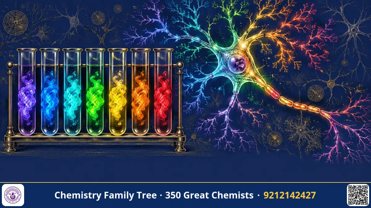
When Osamu Shimomura found GFP and Martin Chalfie showed it worked in other organisms, biologists had one colour: green. Roger Y. Tsien, a chemist at the University of California, San Diego, turned that single colour into a full palette. He explained how the GFP chromophore forms, made mutants that glow blue, cyan, yellow and brighter green, and later helped develop red fluorescent proteins from corals. Before this, he had invented fluorescent dyes that measure calcium inside living cells. For this body of work he shared the 2008 Nobel Prize in Chemistry with Shimomura and Chalfie.
The Scientific Problem Before the Discovery
Two problems stood in front of cell biologists in the 1970s and 1990s.
- Calcium signalling: Ca²⁺ is a universal messenger that triggers muscle contraction, fertilisation, neurotransmitter release and gene expression. But its concentration inside resting cells is only about 100 nM, ten thousand times lower than outside (about 1–2 mM), and it rises only briefly. Existing tools, such as aequorin, were toxic, weak or hard to load.
- One colour: Wild-type GFP was dim and excited mostly by UV; scientists wanted to follow two or more proteins at the same time, or to see protein-protein interactions, which needs different colours.
The Discovery: Step by Step
Part 1: calcium indicators (1980–1985)
- Design of BAPTA (1980): Tsien made a chelator, BAPTA, similar to EGTA but with aromatic rings. It binds Ca²⁺ strongly but is much more selective over Mg²⁺ and is not sensitive to pH changes near physiological values.
- Fluorescent versions: Adding fluorophore groups to the chelator gave Quin-2, then the brighter Fura-2 and Indo-1 (with Grynkiewicz and Poenie), which change their excitation or emission spectra when Ca²⁺ binds.
- The AM-ester trick: The carboxylates of these dyes are masked as acetoxymethyl (AM) esters, which are neutral and cross the membrane. Inside the cell, esterases cleave the esters and trap the charged dye. This made dye loading easy.
Part 2: engineering GFP (1990s)
- Mechanism: Tsien's group showed that GFP's chromophore needs molecular oxygen for the final oxidation (dehydrogenation) step, which explains why it does not fluoresce without O₂.
- Mutations for new colours: By random and rational mutagenesis he changed residues around the chromophore. Replacing Ser65 with Thr (S65T) made GFP brighter, faster to mature and excited at 488 nm, the argon laser line. Changing Tyr66 to His gave blue fluorescent protein (BFP); to Trp gave cyan (CFP). Other changes gave yellow fluorescent protein (YFP).
- Red proteins: The red protein DsRed from a coral (Discosoma) was discovered by others in 1999. Tsien's laboratory re-engineered it into a monomeric form, with a family of "mFruits" such as mCherry, mOrange and mStrawberry.
- Biosensors: Pairs of colours such as CFP and YFP were used as FRET partners to build sensors for calcium (cameleons) and for cAMP and kinase activity.
The Chemistry Behind It
Calcium binding by BAPTA is a simple coordination chemistry equilibrium:
Ca²⁺ + BAPTA⁴⁻ ⇌ [Ca·BAPTA]²⁻ K_d ≈ 10⁻⁷ M
A dissociation constant near 100–200 nM is ideal because it matches the physiological range (100 nM resting to 1 µM active). For a ratio dye like Fura-2, the fluorescence is measured at two excitation wavelengths (340 and 380 nm). Ca²⁺ binding increases emission excited at 340 and decreases that excited at 380. The ratio R = F₃₄₀/F₃₈₀ is then converted to concentration by:
[Ca²⁺] = K_d · β · (R − R_min) / (R_max − R)
Using a ratio removes the effect of dye amount, cell thickness and bleaching. This is a great example of analytical chemistry in biology.
The colour of a fluorescent protein depends on its chromophore and its environment. Changing Tyr66 to His shortens the conjugated system and gives blue light. Adding an additional aromatic ring (as in YFP, with Tyr203 stacking against the chromophore) lowers the excitation energy and moves the emission to yellow. Red proteins extend the conjugation with an additional C=N–C=O unit.
Förster resonance energy transfer (FRET) occurs when the emission spectrum of a donor (CFP) overlaps with the absorption spectrum of an acceptor (YFP). The efficiency depends on distance:
E = 1 / [1 + (r / R₀)⁶] (R₀ ≈ 5 nm for CFP–YFP)
So FRET acts as a "molecular ruler" over 1–10 nm, detecting binding or conformational change.
Applications Today
- Multicolour imaging: Following several proteins at once in living cells.
- Biosensors: Calcium, voltage, pH, ATP and kinase activity in cells and in intact brains (GCaMP sensors are derived from GFP).
- Surgery: Tsien's later work on activatable cell-penetrating peptides, which light up tumours and help surgeons to see their margins.
- Drug discovery: High-throughput screens with fluorescent reporters.
- Education: Coloured bacteria in school biotechnology projects, including Indian synthetic biology competitions.
Exam Corner
- Tsien shared the 2008 Nobel Prize in Chemistry (with Shimomura and Chalfie).
- BAPTA is a Ca²⁺ chelator; Fura-2 is a ratiometric Ca²⁺ dye (340/380 nm).
- AM esters are cell-permeable and trapped by intracellular esterases.
- S65T mutation shifts GFP excitation to 488 nm and brightens it; Y66H gives blue.
- FRET efficiency ∝ 1/r⁶; effective at 1–10 nm.
Common Misconceptions
- "Tsien discovered GFP." No. He improved and extended it; Shimomura discovered it.
- "All fluorescent proteins come from jellyfish." Red ones come from corals.
- "FRET involves emission of a photon by the donor." It is non-radiative energy transfer through dipole coupling.
Quick Revision Box
- BAPTA (1980): selective Ca²⁺ chelator; basis for Quin-2, Fura-2, Indo-1.
- AM ester: masked carboxylates, enter cells, trapped by esterases.
- GFP chromophore needs O₂ to mature.
- Mutants: S65T (bright green), Y66H (blue), CFP, YFP; mCherry from coral protein.
- FRET: E = 1/[1+(r/R₀)⁶].
- Nobel Prize in Chemistry 2008 (shared three ways).
FAQ
Why do cells need low calcium at rest?
A low baseline makes small increases a strong, rapid signal; pumps keep calcium low and prevent precipitation with phosphate.
What does a ratiometric dye mean?
A dye whose signal is measured at two wavelengths, so the ratio removes errors from uneven loading or thickness.
Why did the S65T mutation matter?
It made GFP brighter, more stable and compatible with common laser and filter sets.
Where do red fluorescent proteins come from?
From reef corals and anemones; Tsien's group made them monomeric and brighter.
Published by ABC Chemistry — IIT-JAM | CSIR-NET | GATE Chemistry Coaching. Visit abcchemistry.com

About the chemist
Roger Tsien
1952-2016 · United States · Nobel Chemistry 2008
Engineered fluorescent proteins and calcium probes