The discovery, explained · Chemist no. 142 of 350
Stanford Moore: Counting the Building Blocks of Proteins
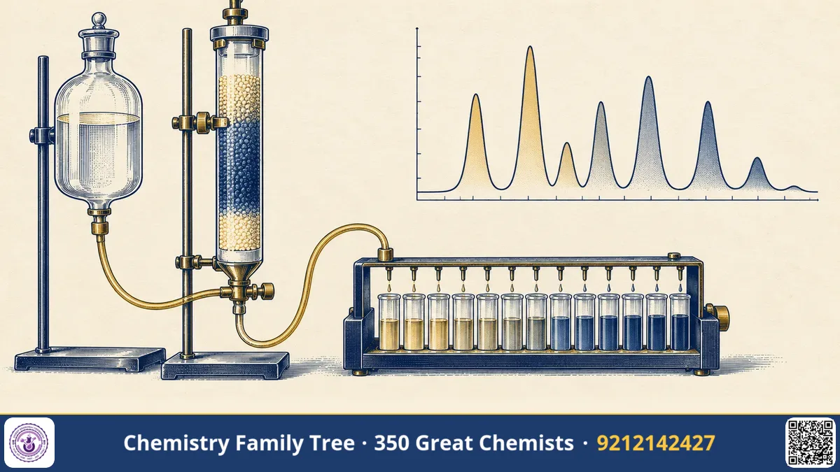
Imagine trying to find out what a protein is made of, when it may contain 20 different amino acids in hundreds of positions. In the 1940s this took weeks of painstaking work and large amounts of material. Stanford Moore and his lifelong colleague William H. Stein of the Rockefeller Institute solved this problem by combining ion-exchange chromatography with the ninhydrin colour reaction, and finally by automating the whole process in the amino acid analyzer. They then used these tools to work out the chemical structure of the enzyme ribonuclease and to identify the amino acids at its active site. For this, Moore and Stein shared one half of the 1972 Nobel Prize in Chemistry; the other half went to Christian Anfinsen.
The Scientific Problem Before the Discovery
Frederick Sanger had shown in the early 1950s that insulin has a precise amino acid sequence, but his methods were slow. Chemists could hydrolyse a protein with acid, but how could the resulting mixture of amino acids be separated and measured? Paper chromatography, introduced by Martin and Synge, helped, but results were only roughly quantitative. Separate spots had to be cut out and measured, which took days and gave errors of 10 percent or more.
A protein chemist needed three things: a clean separation of all twenty amino acids, a sensitive way of detecting them, and a quantitative readout. Without them, comparing proteins, checking purity, or following changes after chemical modification was practically impossible.
The Discovery and Experiments
- Partition chromatography on starch (1948). Moore and Stein first separated amino acids on columns of starch, using solvent mixtures, and collected fractions for analysis with ninhydrin. This already gave a quantitative method, but it was slow.
- Ion-exchange chromatography (1951 onwards). They switched to a sulfonated polystyrene resin (Dowex 50) and eluted with buffers of increasing pH and ionic strength. Amino acids come off the column in a reproducible order.
- Automation (1958). Working with Darrel Spackman, they built a system in which the column effluent is mixed continuously with ninhydrin reagent, heated in a coil, and measured by a colorimeter, with the signal recorded as a chromatogram. A complete amino acid analysis could now be done in a day from a very small sample.
- Applying it to ribonuclease. They determined the amino acid composition and, together with others, the complete sequence of bovine pancreatic ribonuclease A (124 residues, 1960). They also modified the enzyme chemically and showed that histidine residues at positions 12 and 119, along with lysine 41, take part in the active site.
The Chemistry Behind It
Ion-exchange chromatography
The stationary phase is a cation-exchange resin carrying sulfonate groups, R-SO3- H+. At low pH the amino acids are positively charged and stick to the resin by electrostatic attraction:
R-SO3^- H+ + H3N+-CHR-COOH ⇌ R-SO3^- +H3N-CHR-COOH + H+
As the buffer pH rises, the net charge of each amino acid falls, depending on its isoelectric point (pI). Acidic amino acids such as aspartic and glutamic acid, having low pI, come off first. Neutral amino acids follow, and the basic ones such as lysine, histidine and arginine, with high pI, come off last. Hydrophobic interactions between the polystyrene matrix and aromatic side chains also affect the order.
The ninhydrin reaction
Ninhydrin reacts with the free alpha-amino group of amino acids on heating. The amino acid is oxidatively deaminated and decarboxylated, giving an aldehyde, CO2 and ammonia. The reduced ninhydrin (hydrindantin) then condenses with ammonia and another molecule of ninhydrin to give a blue-purple dye known as Ruhemann's purple, which absorbs at 570 nm:
amino acid + 2 ninhydrin → aldehyde + CO2 + Ruhemann's purple (570 nm)
Proline and hydroxyproline, being secondary amines, give a yellow product, measured at 440 nm. The colour intensity is proportional to the amount of amino acid (Beer-Lambert law), so peak area gives quantity.
Hydrolysis
Proteins are hydrolysed with 6 M HCl at about 110 degrees Celsius for 24 hours. Tryptophan is destroyed under these conditions, asparagine and glutamine are converted to aspartic and glutamic acids, and serine and threonine are partly lost. These limitations are well known and are part of exam questions.
Active site chemistry of ribonuclease
Ribonuclease cleaves RNA at the phosphodiester bond through a two-step acid-base mechanism. His-12 acts as a base, accepting a proton from the 2'-hydroxyl group of ribose, while His-119 acts as an acid, donating a proton to the leaving group. A cyclic 2',3'-phosphate intermediate forms and is then hydrolysed. Moore and Stein's modification experiments using iodoacetate, which alkylates histidine, provided early evidence for this picture.
Applications Today
- Protein characterisation. Amino acid analysis remains a standard method to determine the composition and exact concentration of protein and peptide samples, including pharmaceutical peptides.
- Clinical diagnosis. Analysis of amino acids in blood and urine is used to detect inherited metabolic disorders such as phenylketonuria in newborn screening.
- Food and feed industry. Nutritional quality, such as the lysine content in cereals, is checked by amino acid analysis, which is important for India's food quality labs.
- Chromatography in general. Ion-exchange HPLC, water softening and protein purification all use the same principles.
- Enzymology. Chemical modification of residues to find active sites is still taught as a method.
Exam Corner
- Cation-exchange resin (sulfonated polystyrene) separates amino acids by charge: acidic first, basic last, as pH and ionic strength are increased.
- Ninhydrin gives Ruhemann's purple (570 nm) with primary amino acids, and a yellow product (440 nm) with proline.
- Acid hydrolysis (6 M HCl, 110 degrees Celsius, 24 h) destroys tryptophan and converts Asn and Gln to Asp and Glu.
- Ribonuclease A has 124 residues, 4 disulfide bonds; catalytic residues His-12 and His-119.
- The 1972 Nobel Prize in Chemistry was shared by Anfinsen, Moore and Stein.
Common Misconceptions
- "Ninhydrin detects peptide bonds." It detects free alpha-amino groups, so it reacts with free amino acids and N-terminal groups, not with intact peptide bonds.
- "Moore won the Nobel for sequencing insulin." Insulin sequencing was Sanger's work (Nobel 1958). Moore and Stein worked on ribonuclease.
- "Basic amino acids elute first." On a cation exchanger they bind most strongly and elute last.
Quick Revision Box
- Moore and Stein built the first automated amino acid analyzer (1958, with Spackman).
- Resin: Dowex 50, sulfonated polystyrene cation exchanger.
- Detection: ninhydrin, 570 nm (purple) and 440 nm (proline, yellow).
- Elution order tracks isoelectric point: acidic, neutral, then basic.
- Ribonuclease A: 124 amino acids, active-site His-12 and His-119.
- Nobel Prize in Chemistry, 1972 (shared half with Stein).
FAQ
What did Stanford Moore win the Nobel Prize for?
He shared the 1972 Nobel Prize in Chemistry with William Stein for studies on the relationship between chemical structure and catalytic activity of ribonuclease, supported by their analytical methods.
Why use ninhydrin?
It forms a strongly coloured product with amino acids, which allows very sensitive and quantitative detection.
Why do basic amino acids elute last?
They carry a positive charge over a wide pH range and bind strongly to the negatively charged sulfonate groups, so a higher pH or salt concentration is needed to release them.
Is this topic important for exams?
Yes. Amino acid properties, pI, ion-exchange chromatography and ninhydrin are frequent topics in JAM, NET and GATE biochemistry sections.
Published by ABC Chemistry — IIT-JAM | CSIR-NET | GATE Chemistry Coaching. Visit abcchemistry.com
About the chemist
Stanford Moore
1913-1982 · United States · Nobel Chemistry 1972
Ribonuclease structure, amino acid analysis