The discovery, explained · Chemist no. 201 of 350
Stefan Hell and STED Microscopy: Shrinking the Spot of Light
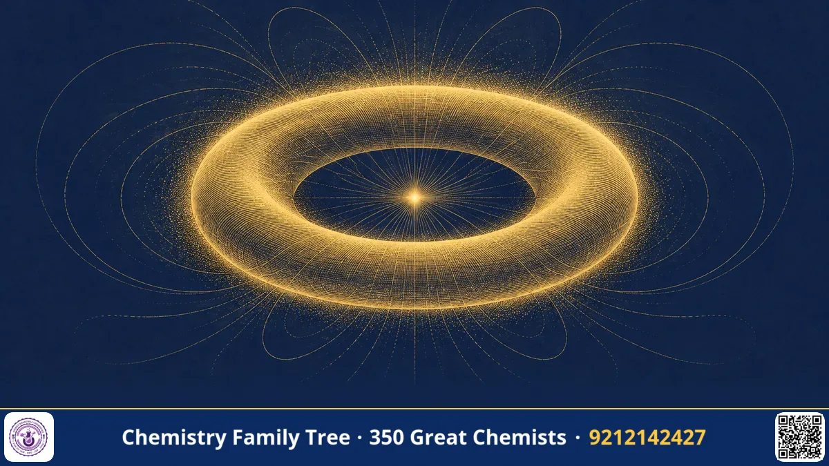
In 1873 Ernst Abbe wrote the law that limited every light microscope: you cannot resolve details closer than about half the wavelength of light. Stefan Hell, a young physicist in Heidelberg, asked a different question in the early 1990s: Abbe's limit applies to how light is focused, but what if the molecules themselves are told when to glow and when not to? He proposed in 1994 and demonstrated in 1999 a method called stimulated emission depletion (STED) microscopy, in which a doughnut-shaped laser beam forces fluorescent molecules around a central spot to go dark, leaving only a tiny glowing region. In 2014 he shared the Nobel Prize in Chemistry with Eric Betzig and William Moerner. This blog explains STED and its chemistry.
The Scientific Problem Before the Discovery
Resolution in a fluorescence microscope is limited by diffraction. When a laser is focused through an objective lens, the focal spot is not a point but a blurred spot, with a width of about λ/(2 NA), around 200-250 nm in the focal plane, and even larger (500-700 nm) along the optical axis. Anything within the spot is excited at the same time and cannot be told apart. Confocal microscopy improved contrast by removing out-of-focus light, but not resolution significantly.
The prevailing opinion, backed by Abbe's theory, was that any optical far-field microscope would be restricted this way. Near-field techniques (NSOM) could do better but only for surfaces, not for the inside of cells. Hell, while a doctoral and postdoctoral researcher, looked for a way to use properties of the fluorescent molecule as a source of resolution.
The Discovery: Step by Step
- The idea (early 1990s). Hell realised that if the fluorescence of molecules in the outer part of the excitation spot could be switched off, only those in the centre would fluoresce, and the effective spot would be narrower than the diffraction limit. A fluorescent molecule can be turned off by stimulated emission.
- Publishing the concept (1994). Together with Jan Wichmann, he published the theory of STED in Optics Letters in 1994, while working at the University of Turku in Finland. He showed that in principle the resolution would have no fixed limit.
- The two-beam design. A first laser pulse (excitation beam) excites fluorophores in a diffraction-limited spot. Immediately afterwards, a second, red-shifted pulse (the STED beam), shaped like a doughnut with zero intensity at the centre, forces excited molecules at the periphery to return to the ground state by stimulated emission, before they can fluoresce. Only molecules in the centre remain, and they emit fluorescence normally.
- Experimental proof (1999). At the Max Planck Institute for Biophysical Chemistry in Göttingen, Hell and Thomas Klar demonstrated the method, first on test samples and soon on biological samples such as yeast cells and bacteria, reaching resolutions of about 100 nm. Later versions reached 20-30 nm in the focal plane.
- Scanning and imaging. The small spot is scanned over the sample, point by point, to build an image. Further development led to STED in living cells and to video-rate imaging.
- MINFLUX (2016-2017). Hell's group later combined ideas from STED and single-molecule localisation in MINFLUX, in which a doughnut-shaped excitation beam locates individual molecules with a precision of a few nanometres using few photons.
The Chemistry Behind It
Stimulated emission was predicted by Einstein in 1917 and underlies the laser. A molecule in the excited state S₁ can return to the ground state S₀ in several ways. Without interference, it emits a photon spontaneously (fluorescence, with a lifetime of a few nanoseconds). If a photon of the right wavelength arrives, it can stimulate the molecule to emit a second photon with the same wavelength, phase and direction:
S₀ + hν(exc) → S₁
S₁ → S₀ + hν(fl)              (spontaneous emission, fluorescence)
S₁ + hν(STED) → S₀ + 2hν(STED) (stimulated emission, no fluorescence detected)
The STED wavelength is chosen at the red edge of the emission spectrum, so it does not excite the dye and is easy to separate from fluorescence in the detector. Stimulated emission competes with fluorescence. The fraction of excited molecules that remain fluorescent after the STED pulse falls exponentially with the STED intensity I. The new resolution is given by:
d ≈ λ / (2 NA √(1 + I/I_s))
where I_s is the saturation intensity of the dye (the intensity at which half of the excited molecules are depleted). When I is much greater than I_s, d becomes small; in principle the resolution is limited only by how much STED light the sample and dye can tolerate before photobleaching or damage. Dyes for STED must therefore be photostable, with a large Stokes shift and efficient stimulated emission. The doughnut shape is produced with a phase plate that gives the beam a helical phase, so that destructive interference creates a zero at the centre.
Applications Today
- Neuroscience. STED has allowed scientists to image the shape of dendritic spines and synapses in living brain tissue, and to watch vesicle movements in neurons.
- Cell biology. It has been used to study the organisation of mitochondria, the endoplasmic reticulum, the cytoskeleton and nuclear pore complexes.
- Virus research. Researchers use STED to see how viruses assemble and enter cells.
- Chemistry of dyes. The technique spurred the design of new fluorophores and labels, including self-labelling protein tags and live-cell dyes.
- Commercial instruments. STED microscopes are now sold by several manufacturers and are used in research institutes worldwide, including facilities in India.
Exam Corner
- Nobel Prize in Chemistry 2014: Betzig, Hell and Moerner, for super-resolved fluorescence microscopy.
- STED = stimulated emission depletion; theory published in 1994, first experiments in 1999.
- Abbe limit: d = λ/(2 NA); STED modifies this to d = λ/(2 NA √(1 + I/I_s)).
- Stimulated emission produces a photon identical to the stimulating photon; Einstein introduced the concept in 1917.
- STED beam has a doughnut profile with zero intensity at the centre.
Common Misconceptions
- "STED uses a smaller wavelength to see better." It uses visible light; the resolution gain comes from controlling the molecular states.
- "Stimulated emission is the same as fluorescence." Fluorescence is spontaneous, while stimulated emission is triggered by an incoming photon.
- "STED is a scanning electron method." It is purely optical and works on fluorescently labelled samples, including living cells.
Quick Revision Box
- STED proposed by Hell in 1994; demonstrated in 1999.
- Excitation beam plus doughnut-shaped depletion beam.
- Molecules at the periphery are returned to S₀ by stimulated emission.
- d ≈ λ / (2 NA √(1 + I/I_s)).
- Dyes need photostability and strong stimulated emission.
- Nobel Prize in Chemistry 2014.
FAQ
How does STED differ from PALM?
STED shrinks the excitation spot using a patterned laser, working point by point, while PALM images sparse single molecules and calculates their positions.
Why a doughnut beam?
The zero-intensity centre leaves molecules there undisturbed, while molecules in the ring around it are switched off.
Does STED damage cells?
High STED intensity can cause photodamage, so researchers optimise dyes, wavelength and intensity for live-cell work.
What is MINFLUX?
A later method from Hell's laboratory that locates single molecules with precision of a few nanometres using a doughnut-shaped excitation beam.
Published by ABC Chemistry — IIT-JAM | CSIR-NET | GATE Chemistry Coaching. Visit abcchemistry.com

About the chemist
Stefan Hell
1962- · Germany · Nobel Chemistry 2014
STED super-resolution microscopy