The discovery, explained · Chemist no. 192 of 350
Thomas Steitz and the Ribosome: The Day We Learned That RNA Makes Proteins
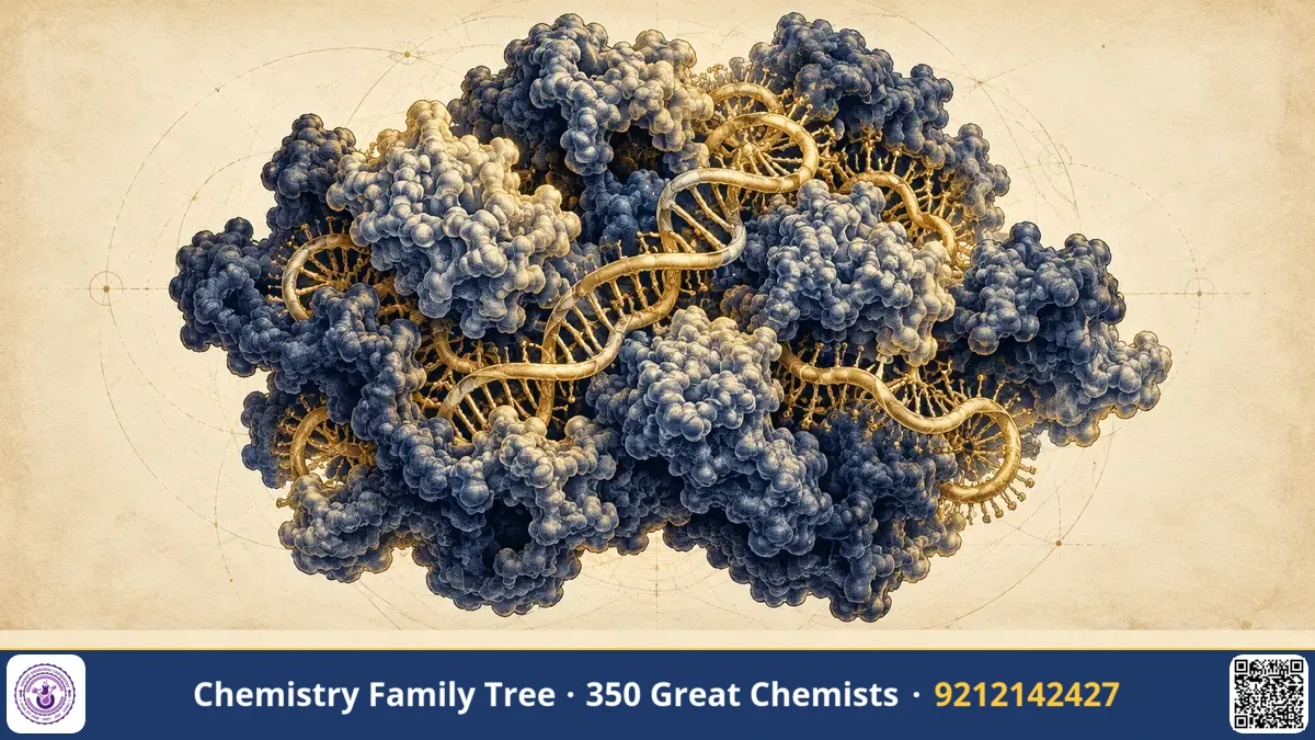
Every protein in your body, from haemoglobin to the enzymes digesting your lunch, was assembled by a molecular machine called the ribosome. For decades chemists believed that this machine worked like most enzymes, with protein side chains doing the catalytic work. In 2000, the group of Thomas A. Steitz at Yale University published an atomic-resolution picture of the large ribosomal subunit and showed something astonishing: the chemistry of peptide-bond formation is catalysed by RNA. The ribosome is a ribozyme. For this work, shared with Ada Yonath and Venkatraman Ramakrishnan, Steitz received the Nobel Prize in Chemistry in 2009. This post explains the problem, the experiment, the chemistry and the exam points you should remember.
The Scientific Problem Before the Discovery
By the 1960s the broad outline of protein synthesis was known. DNA is transcribed to messenger RNA (mRNA); transfer RNA (tRNA) carries amino acids; and ribosomes read the mRNA in groups of three bases (codons) and join the amino acids in order. The ribosome itself was known to contain about two-thirds RNA (ribosomal RNA, rRNA) and one-third protein by mass. In bacteria the complete 70S ribosome is made of a small 30S subunit and a large 50S subunit; in eukaryotes the 80S ribosome has 40S and 60S subunits. (Svedberg values are sedimentation coefficients, so they do not add up.)
Two big questions remained. First, what does the ribosome look like at atomic resolution? Second, which part of it actually forms the peptide bond? Because ribosomes contain dozens of proteins, most scientists assumed one of them was the "peptidyl transferase". In 1982 Thomas Cech and Sidney Altman had shown that RNA can be catalytic (ribozymes), and Harry Noller later showed that peptidyl transferase activity survived harsh protein-removing treatments, hinting at RNA catalysis. But hints are not proof. The ribosome has about 4,500 nucleotides of rRNA in the large subunit alone, so crystallising it and solving its structure looked almost impossible.
The Discovery: Step by Step
- Getting crystals. Ada Yonath had grown the first ribosome crystals around 1980. Steitz, working with Peter Moore at Yale, chose the large subunit of the salt-loving archaeon Haloarcula marismortui, which gave well-ordered crystals.
- X-ray diffraction. Crystals were exposed to intense synchrotron X-rays. The diffraction spots give amplitudes, but the phases are lost, which is the classic "phase problem".
- Solving phases. The team used heavy-atom derivatives, such as clusters containing tungsten or other heavy metals, together with lower-resolution electron-microscopy maps, to obtain phases, then built an atomic model into the electron-density map.
- The 2.4 Å structure (2000). The paper from Steitz's laboratory, with Nenad Ban, Poul Nissen, Jeffrey Hansen and Peter Moore, reported the 50S subunit at 2.4 Å resolution. At this resolution individual nucleotides and amino-acid side chains are visible.
- Locating the active site. By soaking in a transition-state analogue of the peptidyl transferase reaction, the team marked exactly where peptide-bond formation occurs. Surrounding it were only RNA helices. No protein lay within about 18 Å of the catalytic centre.
The conclusion was clear: the peptidyl transferase centre is built of 23S-type rRNA, and proteins sit on the surface, stabilising the fold. Steitz summarised this as the ribosome being a ribozyme.
The Chemistry Behind It
The reaction catalysed is a nucleophilic acyl substitution. The growing peptide is attached as an ester to the 3'-OH of the tRNA in the P site, and the incoming aminoacyl-tRNA sits in the A site. The α-amino group of the A-site amino acid attacks the carbonyl carbon of the ester:
Peptidyl(n)-tRNA(P site) + aminoacyl-tRNA(A site) → peptidyl(n+1)-tRNA(A site) + deacylated tRNA(P site)

R-CO-O-tRNA + H2N-CHR'-CO-O-tRNA' → R-CO-NH-CHR'-CO-O-tRNA' + HO-tRNA
Notice that the ester bond to the old tRNA is broken while a stable amide (peptide) bond forms. The ribosome does not use ATP for this step; the energy comes from the high-energy aminoacyl ester bond made earlier by aminoacyl-tRNA synthetase.
How does RNA speed up this reaction? The structure suggested that the ribosome works mainly as an entropy trap. It positions the two substrates precisely and in the right orientation, and it excludes water. A universally conserved adenine in the active site (A2486 in the H. marismortui numbering; A2451 in E. coli) is placed near the attacking amine. Later work indicated that the 2'-OH of the P-site tRNA's terminal adenosine takes part in a proton shuttle that helps the reaction proceed. Detailed mechanism is still researched, but the central message has not changed: the catalyst is RNA.
This supports the "RNA world" hypothesis, the idea that early life used RNA for both information storage and catalysis. A catalytic core made of RNA in every living ribosome looks like a molecular fossil from that era.
Applications Today
- Antibiotic design. Many antibiotics work by binding bacterial ribosomes: erythromycin and other macrolides block the exit tunnel, chloramphenicol sits in the peptidyl transferase centre, tetracycline binds the 30S subunit near the A site, and streptomycin causes misreading. Steitz's team solved structures of several antibiotics bound to the 50S subunit, showing how they bind and how resistance mutations (for example methylation of rRNA) block them. Steitz co-founded a company, Rib-X Pharmaceuticals, to use ribosome structures to design new antibiotics.
- Antimicrobial resistance. In India, where antibiotic resistance is a serious public-health concern, structure-guided drug design is a key route to new medicines.
- Selectivity. Bacterial 70S ribosomes differ from human 80S ribosomes, so drugs can attack bacteria while sparing our cells.
- Synthetic biology. Understanding the peptidyl transferase centre helps scientists engineer ribosomes that incorporate unnatural amino acids.
Exam Corner
- The 2009 Nobel Prize in Chemistry went to Venkatraman Ramakrishnan, Thomas Steitz and Ada Yonath for studies of the structure and function of the ribosome.
- Steitz's 2000 structure was of the 50S (large) subunit of Haloarcula marismortui at 2.4 Å, solved by X-ray crystallography.
- The peptidyl transferase centre is formed by rRNA, so the ribosome is a ribozyme. Cech and Altman received the 1989 Nobel Prize in Chemistry for catalytic RNA.
- Peptide bond formation is nucleophilic attack of the A-site α-amino group on the P-site ester carbonyl; no ATP is used in this step.
- Prokaryotic ribosome: 70S = 30S + 50S; eukaryotic: 80S = 40S + 60S. The mRNA is decoded by the small subunit, and the peptide bond is made in the large subunit.
Common Misconceptions
- "Ribosomal proteins make the peptide bond." No. The active site is RNA-only; proteins stabilise the structure and help the nascent chain.
- "Only Steitz solved the ribosome." Three independent groups solved major parts: Steitz (50S), Yonath (50S and 30S work) and Ramakrishnan (30S). Their results complemented each other.
- "Ribosomes need ATP to form each peptide bond." The bond-forming step does not need ATP. GTP is used by elongation factors for translocation and delivery of tRNA, and ATP is used earlier to charge tRNAs.
Quick Revision Box
- Steitz: Nobel Prize in Chemistry 2009 (shared with Yonath and Ramakrishnan).
- Structure: 50S subunit of H. marismortui, 2.4 Å, published in 2000.
- Key finding: no protein near the active site, so the ribosome is a ribozyme.
- Reaction: aminolysis of peptidyl-tRNA ester, forming an amide bond.
- Method: X-ray crystallography with heavy-atom phasing.
- Application: structure-based antibiotic design.
FAQ
What is a ribozyme?
A ribozyme is an RNA molecule that acts as a catalyst. Examples include self-splicing introns, RNase P and the peptidyl transferase centre of the ribosome.
Why did the ribosome take so long to crystallise?
It is huge, flexible and made of many components, and crystals need every molecule to sit in the same orientation. Years of work on stability, purification and freezing were needed.
What does 2.4 Å resolution mean?
One Ångström is 10⁻¹⁰ m. A resolution of 2.4 Å means features about that far apart can be distinguished, enough to see individual atoms of nucleotides and the sites where antibiotics bind.
Is this topic important for CSIR-NET and GATE?
Yes. Protein synthesis, ribozymes, X-ray crystallography and antibiotic action on the ribosome appear regularly in the Life Sciences and Chemical Sciences papers, and the Nobel Prize list is a popular source of questions.
Published by ABC Chemistry — IIT-JAM | CSIR-NET | GATE Chemistry Coaching. Visit abcchemistry.com
About the chemist
Thomas A. Steitz
1940-2018 · United States · Nobel Chemistry 2009
Atomic structure of the ribosome