The discovery, explained · Chemist no. 191 of 350
Venki Ramakrishnan and the Ribosome: The Atomic Structure of the Protein Factory
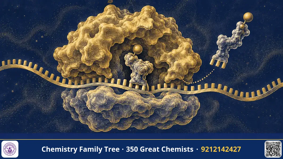
Every protein in your body, from haemoglobin to the enzymes that digest your food, is built by a molecular machine called the ribosome. It reads the sequence of messenger RNA (mRNA) and joins amino acids in the correct order, with astonishing accuracy. For decades the ribosome was too large and complex to be seen at atomic detail. In 1999–2000, a group led by Indian-born scientist Venkatraman (Venki) Ramakrishnan at the MRC Laboratory of Molecular Biology in Cambridge solved the structure of the 30S ribosomal subunit, and showed how it decodes the genetic message and how antibiotics disturb it.
In 2009 Ramakrishnan shared the Nobel Prize in Chemistry with Thomas A. Steitz and Ada E. Yonath "for studies of the structure and function of the ribosome".
The Scientific Problem Before the Discovery
By 1970 biochemists knew the outline of translation: mRNA codons are read by transfer RNAs (tRNAs) carrying amino acids, and the ribosome is the site where peptide bonds form. Bacterial ribosomes (70S) consist of a small subunit (30S: 16S rRNA plus about 20 proteins) and a large subunit (50S: 23S and 5S rRNA plus about 30 proteins). But important questions remained:
- How does the small subunit choose the correct tRNA? The codon–anticodon pairing energy is small, which should give an error rate of about 1 in 10 or 100, yet the real error rate of translation is about 1 in 1,000 to 10,000.
- Which part is the catalyst, protein or RNA? (Later shown to be rRNA: the ribosome is a ribozyme.)
- How do antibiotics such as streptomycin, tetracycline and paromomycin kill bacteria without affecting human ribosomes?
Crystallising a ribosome was extremely difficult: it has over a million atoms, is flexible and is hard to purify. Many experts in the 1980s thought it impossible.
The Discovery: Step by Step
- Choose a stable source: The team used ribosomes from the heat-loving bacterium Thermus thermophilus, which lives in hot springs, since its proteins are more stable and form better crystals.
- Crystallise the 30S subunit: Crystals diffracted X-rays only weakly, but with synchrotron radiation and many improvements the team obtained data to about 3 Å resolution.
- Solve the phase problem: They used heavy-atom clusters and anomalous scattering to obtain phases, then built the atomic model of 16S rRNA and 20 proteins (published in Nature, 2000).
- Add ligands: Further crystals were soaked in antibiotics (paromomycin, streptomycin, spectinomycin) to see where they bind.
- Add mRNA and tRNA fragments: Complexes with an anticodon stem-loop and a short mRNA showed what happens during codon recognition. Later work solved whole 70S ribosomes with tRNAs and with elongation factors.
The Chemistry Behind It
The structure explains accurate decoding through induced fit and kinetic proofreading. In the decoding centre of the 30S subunit, two universally conserved adenines of 16S rRNA, A1492 and A1493, flip out of helix 44 and press on the minor groove of the first two base pairs formed between the mRNA codon and the tRNA anticodon. They monitor the shape of the codon–anticodon helix, which is correct only for Watson–Crick pairs (A–U, G–C). Hydrogen bonds between the 2′-OH and the bases are formed, as in the A-minor interaction:
Correct (cognate) tRNA → full A-minor contacts → 30S domain closure → GTP hydrolysis by EF-Tu → accommodation
Wrong (near-cognate) tRNA → poor contacts → no stable closure → tRNA rejected
The binding energy difference between right and wrong pairings (ΔΔG of roughly 3 kcal/mol) alone would give an error rate near 10⁻²:
error ≈ exp(−ΔΔG / RT) (~10⁻² for ΔΔG ≈ 2.7 kcal/mol at 310 K)
Cells need a better accuracy; they achieve it by two separate selection steps (initial selection and proofreading) with a GTP hydrolysis step between them, which increases discrimination to about 10⁻⁴.
Antibiotics exploit the same structure. Paromomycin, an aminoglycoside, binds in helix 44 near A1492/A1493 and forces them into the flipped-out state even with a wrong tRNA, causing misreading. Streptomycin binds near the decoding centre and stabilises a closed form, also increasing errors. Tetracycline blocks the A site so that aminoacyl-tRNA cannot enter. Because bacterial and human ribosomes differ in details of the rRNA and proteins, these drugs can be selective.
Applications Today
- Antibiotic design: Structures guide the design of drugs that overcome resistance, an urgent need in India where antibiotic resistance is high.
- Understanding resistance: Methylation of A1408 or A2058 of rRNA changes drug binding; structures show how.
- Synthetic biology: Engineered ribosomes can incorporate unnatural amino acids.
- Cancer and genetic diseases: Ribosomopathies and the role of ribosome heterogeneity are now studied in human cells.
- Cryo-EM: Later work by the same laboratory used cryo-electron microscopy to solve human and mitochondrial ribosomes.
Exam Corner
- Ramakrishnan, Steitz and Yonath shared the Nobel Prize in Chemistry 2009 for the structure and function of the ribosome.
- Bacterial ribosome 70S = 30S (16S rRNA + ~20 proteins) + 50S (23S, 5S rRNA + ~30 proteins).
- Decoding centre: A1492 and A1493 of 16S rRNA monitor codon–anticodon geometry.
- Peptide bond formation is catalysed by rRNA (23S), so the ribosome is a ribozyme (Steitz's work on the 50S subunit).
- Aminoglycosides cause misreading; tetracycline blocks the A site; macrolides block the exit tunnel.
Common Misconceptions
- "The ribosome is mostly protein." By mass it is about two-thirds RNA, and the catalytic and decoding sites are formed by RNA.
- "Ramakrishnan solved the whole ribosome alone." The 30S work was his; Steitz and Yonath solved the 50S and 30S in parallel, and many co-workers took part.
- "Human and bacterial ribosomes are identical." They differ in size (80S versus 70S) and in rRNA details.
Quick Revision Box
- 30S subunit structure from Thermus thermophilus, published 2000.
- Decoding by A1492/A1493 flipping and A-minor contacts.
- Accuracy about 10⁻⁴ via induced fit plus proofreading.
- Paromomycin and streptomycin induce misreading.
- The ribosome is a ribozyme; rRNA forms the peptidyl transferase centre.
- Nobel Prize in Chemistry 2009, shared three ways.
FAQ
What is the Svedberg unit in 30S and 50S?
S is a sedimentation coefficient measured in centrifugation; it depends on size and shape, so 30S plus 50S gives 70S, not 80S.
Why is X-ray crystallography used for the ribosome?
It gave the first atomic-resolution structures; today cryo-EM is also widely used.
How does the ribosome make peptide bonds?
The amino group of the A-site aminoacyl-tRNA attacks the ester carbonyl of the P-site peptidyl-tRNA, and the rRNA positions the substrates and stabilises the transition state.
Why are ribosomes targets for antibiotics?
Bacteria need ribosomes to make proteins, and the differences from human ribosomes allow selective inhibition.
Published by ABC Chemistry — IIT-JAM | CSIR-NET | GATE Chemistry Coaching. Visit abcchemistry.com

About the chemist
Venkatraman Ramakrishnan
1952- · United Kingdom · Nobel Chemistry 2009
Structure of the ribosome