The discovery, explained · Chemist no. 150 of 350
Aaron Klug: Seeing the Shapes of Viruses and Gene-Controlling Proteins
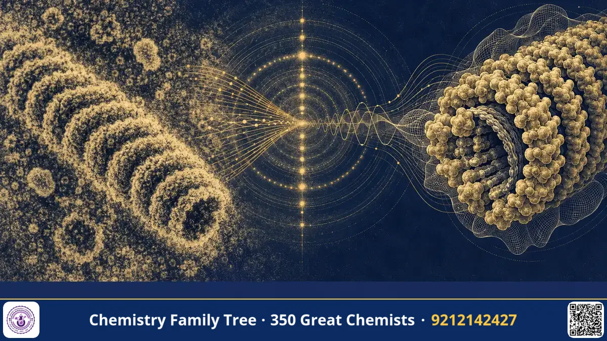
An electron microscope can magnify a virus a hundred thousand times, but the picture it gives is flat, a shadow of a three-dimensional object. How can we turn such flat images into a true 3D model? Aaron Klug (1926-2018) found the answer by combining electron microscopy with the mathematics of Fourier transforms and the ideas of X-ray crystallography. He used it to study viruses, chromatin and protein-nucleic acid complexes, and he discovered a new structural motif - the zinc finger - which helps proteins recognise DNA. For this work he received the 1982 Nobel Prize in Chemistry for the development of crystallographic electron microscopy and his structural elucidation of biologically important nucleic acid-protein complexes.
The Scientific Problem Before the Discovery
X-ray crystallography can solve the structures of molecules which form good crystals, but many biological structures - viruses, ribosomes, filaments and membranes - are difficult or impossible to crystallise. Electron microscopy can image them directly, but biological samples are damaged by the electron beam, and they must be viewed in vacuum. Also, a micrograph is a projection: the density of all the atoms along the beam direction is added up, so depth information is lost.
There is also a mathematical issue. In X-ray diffraction, we measure the amplitudes of diffracted waves but not their phases, which is the "phase problem". In the electron microscope, a lens forms an image, and the image contains both amplitude and phase information. If this information could be combined from many views, a three-dimensional structure could be computed.
The Discovery and Experiments
- Tobacco mosaic virus and spherical viruses. At Birkbeck College, London, and later at Cambridge, Klug continued the X-ray work on viruses started with Rosalind Franklin, whose collaborator he was. He studied the structure of tobacco mosaic virus (TMV), a helical rod made of coat-protein subunits wound around an RNA strand. With Donald Caspar he developed the theory of quasi-equivalence to explain the architecture of spherical viruses built from many identical subunits.
- Optical diffraction of micrographs. Klug used a simple optical bench with a laser to produce the diffraction pattern of an electron micrograph. This tells how regular the image is and allows the best images to be chosen.
- Three-dimensional reconstruction (1968). With David DeRosier, Klug showed how a set of 2D projections from different angles can be combined to compute a 3D map, using the Fourier transform. They first applied it to the tail of bacteriophage T4.
- Nucleosome structure. In the 1970s, Klug's team studied chromatin and showed that DNA wraps around a core of histone proteins, forming the nucleosome core particle with about 146 base pairs of DNA, wound in nearly two turns around a histone octamer.
- Zinc fingers (1985). Klug and colleagues, studying the transcription factor TFIIIA from Xenopus (frog) eggs, found that the protein contains repeating units, each of around 30 amino acids, which bind a zinc ion. They proposed that each unit forms a finger-like domain that fits into the major groove of DNA.
The Chemistry Behind It
Fourier transform and the projection theorem
Any periodic function can be written as a sum of waves; the Fourier transform converts between real space (the image) and reciprocal space (the diffraction pattern). The central section theorem states that the Fourier transform of a 2D projection of an object is a central slice of the 3D Fourier transform of the object, taken perpendicular to the viewing direction. If we collect projections from many angles, the central slices fill the 3D Fourier space, and an inverse Fourier transform gives the 3D structure:
ρ(x,y,z) = Σ F(h,k,l) exp[-2πi(hx + ky + lz)]
where ρ is the electron density and F(h,k,l) is the structure factor, with both amplitude and phase. In electron microscopy, the image gives both the amplitude and the phase, so the phase problem disappears.
Helical symmetry
For helical objects like TMV, one micrograph shows the object from several angles, thanks to the symmetry, so that a single image can be enough for a 3D reconstruction. For a helix with u subunits in t turns, the repeat is called the helical repeat, and the diffraction pattern lies on layer lines described by Bessel functions (Cochran, Crick and Vand).
Zinc finger chemistry
The classical Cys2His2 zinc finger follows the pattern:
Tyr/Phe-X-Cys-X(2-4)-Cys-X(3)-Phe-X(5)-Leu-X(2)-His-X(3-5)-His
Two cysteines and two histidines are bound tetrahedrally to a Zn2+ ion, folding a short beta hairpin and an alpha helix into a compact fold (a ββα fold). The helix goes into the major groove of DNA, and its amino acids make contacts with about three base pairs. Chaining three or more fingers lets a protein read a longer DNA sequence, with each finger recognising its triplet. This is a good example of coordination chemistry in biology: Zn2+ (d10) is redox-inactive and has a stable tetrahedral geometry.
Nucleosome
Histones (H2A, H2B, H3, H4) are rich in the basic amino acids lysine and arginine. They interact with the negatively charged phosphate groups of DNA by electrostatic attractions and hydrogen bonds. Two copies of each of the four histones make the octamer, around which about 146 base pairs of DNA are wrapped in 1.65 left-handed superhelical turns.
Applications Today
- Cryo-electron microscopy. The modern technique of cryo-EM, which won the 2017 Nobel Prize in Chemistry (Dubochet, Frank and Henderson), uses the same principles of image processing and 3D reconstruction from many 2D projections. It is used to visualise ribosomes, membrane proteins and viruses, including the spike protein of SARS-CoV-2.
- Medical imaging. The same mathematics underlies computed tomography (CT) scanners, which reconstruct body images from X-ray projections.
- Genome engineering. Zinc finger nucleases, made by linking zinc fingers to a DNA-cutting enzyme domain, were an early gene editing tool, before TALENs and CRISPR-Cas9.
- Drug discovery. Structural biology of viral proteins and receptors is used to design drugs and vaccines.
- Epigenetics. The nucleosome structure is central to modifications of histones that regulate genes.
Exam Corner
- Klug received the 1982 Nobel Prize in Chemistry (sole award) for crystallographic electron microscopy.
- The nucleosome core particle has about 146 base pairs of DNA around a histone octamer (two each of H2A, H2B, H3, H4); H1 is the linker histone.
- Cys2His2 zinc fingers bind Zn2+ tetrahedrally and recognise about three base pairs each.
- Central section theorem: the Fourier transform of a projection is a central slice of the 3D transform.
- TMV is a helical RNA virus with 2130 coat protein subunits.
Common Misconceptions
- "Zinc fingers are enzymes." They are DNA-binding domains; zinc has a structural role.
- "Klug invented the electron microscope." Ernst Ruska built the first one; Klug developed how to turn the images into 3D structures.
- "Klug won the prize in Physics or Medicine." It was the Nobel Prize in Chemistry.
Quick Revision Box
- Method: 2D electron micrographs plus Fourier transforms give 3D structure.
- First applied to bacteriophage T4 tail (DeRosier and Klug, 1968).
- Nucleosome: 146 bp DNA on a histone octamer.
- Zinc finger: Cys2His2, Zn2+, ββα fold, found in TFIIIA.
- Quasi-equivalence theory with Caspar for virus capsids.
- Nobel Prize in Chemistry, 1982.
FAQ
What is crystallographic electron microscopy?
It is the use of electron micrographs of regular objects and Fourier methods, borrowed from X-ray crystallography, to compute three-dimensional structures.
What is a zinc finger?
It is a small protein domain that uses a zinc ion held by cysteine and histidine side chains to fold into a shape that binds DNA.
Did Klug work with Rosalind Franklin?
Yes. He joined her group at Birkbeck College and continued the virus work after her death in 1958.
How is it useful in exams?
Questions on nucleosome structure, DNA-binding motifs, zinc coordination and Fourier methods in structure determination appear in chemistry and life-science papers.
Published by ABC Chemistry — IIT-JAM | CSIR-NET | GATE Chemistry Coaching. Visit abcchemistry.com

About the chemist
Aaron Klug
1926-2018 · United Kingdom · Nobel Chemistry 1982
Crystallographic electron microscopy