The discovery, explained · Chemist no. 212 of 350
George P. Smith and Phage Display: Linking Proteins to Their Genes
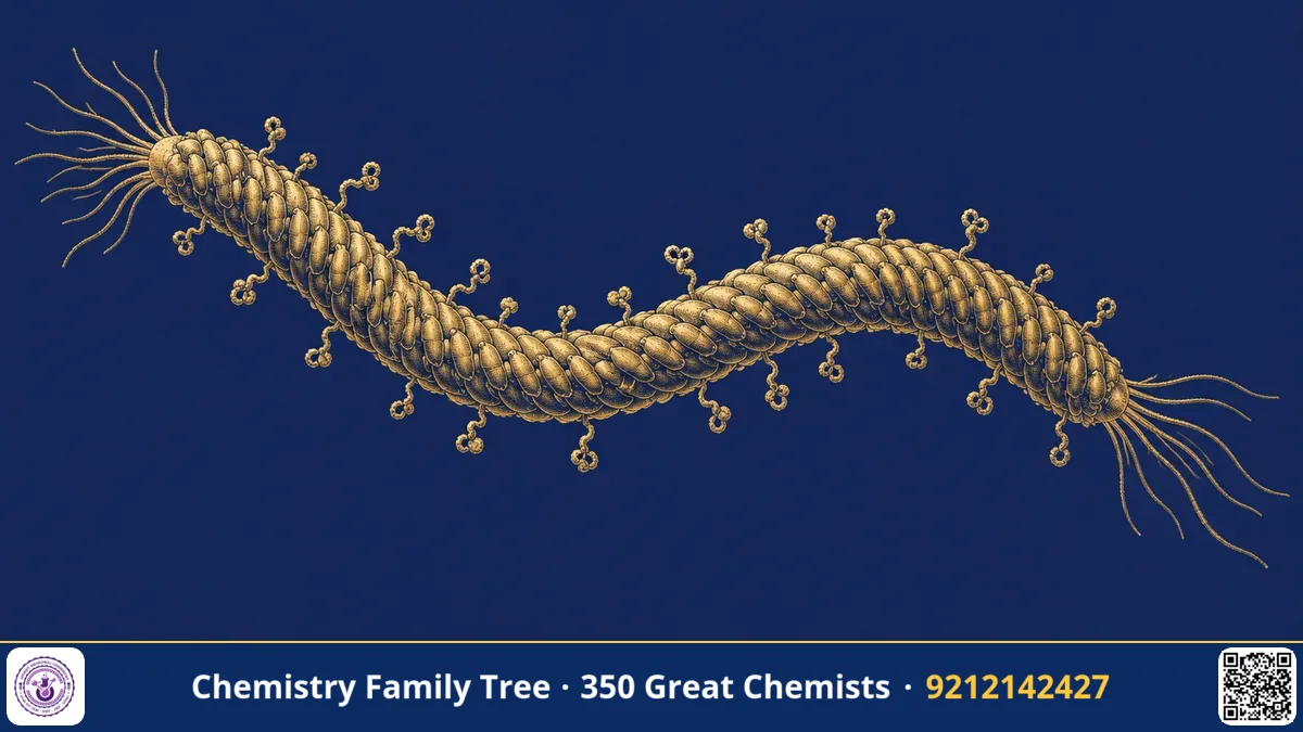
Suppose you want to find a peptide or antibody that binds tightly to a particular target, such as a cancer protein. Billions of possible sequences exist. How do you find the best one? In 1985, George P. Smith of the University of Missouri solved a key part of this puzzle by displaying foreign peptides on the surface of a virus that infects bacteria, a bacteriophage. Because each virus particle carries the gene for the peptide it displays, the physical molecule and its genetic information are linked. This technique, phage display, earned Smith half of the 2018 Nobel Prize in Chemistry; the other half went to Sir Gregory Winter. (Frances Arnold received the other half of the prize for directed evolution of enzymes.)
The Scientific Problem Before the Discovery
In the 1980s, recombinant DNA technology allowed scientists to clone genes and produce proteins. But screening was slow. If you had a large collection of gene variants, you had to express each one and test its protein separately. There was no easy way to select a protein for a property such as binding and then immediately know which gene encoded it.
Nature solves this problem in the immune system and in evolution, where a phenotype (the function of a molecule) is linked to the genotype (the sequence). Smith's insight was to create an artificial link between genotype and phenotype in a test tube, allowing Darwinian selection to work on proteins in the laboratory.
The Discovery: Step by Step
Step 1: Choose the virus
Smith worked with the filamentous bacteriophage f1 (closely related to M13) that infects E. coli. These phages are long thin rods whose coat is made mostly of thousands of copies of a major coat protein and a few copies of minor coat proteins such as pIII, located at one tip. The phage genome is a circular single-stranded DNA.
Step 2: Insert a foreign gene
Smith inserted a fragment of DNA encoding a piece of the restriction enzyme EcoRI into the gene for the minor coat protein pIII. When the phage was assembled, the foreign peptide was fused to pIII and displayed on the surface of the phage, while the DNA for it was inside the same particle.
Step 3: Prove it is displayed
He showed that antibodies against the EcoRI fragment could bind to these phage particles and that the phage could be separated from ordinary phage using the antibody. This was the proof of principle: a protein fragment on the surface, with its gene inside, can be pulled out by affinity.
Step 4: Biopanning
Later, Smith and others made libraries of billions of phages, each displaying a different random peptide. The selection cycle, called biopanning, works as follows:
- Immobilise the target (for example an antibody or a receptor) on a surface.
- Add the phage library; binders stick.
- Wash away non-binders.
- Elute the bound phages (for example by changing pH).
- Infect E. coli with the eluted phage to amplify them.
- Repeat for 3 to 5 rounds, enriching the best binders, then sequence the DNA to read the peptide.
The Chemistry Behind It
- Genotype-phenotype link: Each phage particle packages the DNA that encodes the displayed protein.
- Affinity chemistry: Binding depends on non-covalent interactions: hydrogen bonds, electrostatics, hydrophobic contacts and shape complementarity. The dissociation constant Kd measures strength; lower Kd means tighter binding.
- Fusion proteins: The foreign sequence is encoded in-frame with pIII, so the translated product is a single polypeptide chain.
- Enrichment: If a binder is present at 1 in 10⁹ initially and each round enriches it by 1000-fold, it dominates after three rounds.
- Standard genetic code and PCR: After selection, the sequence of the peptide is read from the DNA, a straightforward use of molecular biology.
Target-binding step: Phage-peptide + Target ⇌ Phage-peptide·Target Kd = [Phage][Target] / [Complex]
Applications Today
- Therapeutic antibodies: Gregory Winter used phage display to evolve human antibodies; the first drug of this kind, adalimumab (Humira), treats rheumatoid arthritis and other inflammatory diseases.
- Peptide drug discovery: Finding peptides that bind receptors, enzymes or cell surfaces.
- Epitope mapping: Finding which part of an antigen an antibody recognises, useful for vaccine design.
- Diagnostics: Identifying binders for disease markers.
- Materials science: Peptides selected to bind minerals or nanoparticles.
Exam Corner
- What is phage display? Display of peptides or proteins on the surface of a phage as fusions to a coat protein, with the encoding gene inside.
- Which coat protein was used by Smith? Minor coat protein pIII of filamentous phage.
- What is biopanning? Iterative affinity selection of binders from a phage library.
- Who shared the 2018 Chemistry Nobel? Frances Arnold (one half), George Smith and Gregory Winter (other half).
- Phage genome type: single-stranded circular DNA (for filamentous phages).
Common Misconceptions
- "Phages infect human cells." Bacteriophages infect only bacteria.
- "Phage display makes proteins from scratch." It selects from existing libraries.
- "The Nobel Prize 2018 was only about phage display." It also recognised Arnold's directed evolution of enzymes.
Quick Revision Box
- Smith introduced phage display in 1985.
- Foreign peptide fused to phage coat protein pIII.
- Gene inside, protein outside: genotype-phenotype link.
- Biopanning: bind, wash, elute, amplify, repeat.
- Led to human therapeutic antibodies such as adalimumab.
- Nobel Prize in Chemistry 2018 (half shared by Smith and Winter).
FAQ
Why do we amplify phages in E. coli?
Phages replicate inside bacteria, so infecting bacteria gives millions of copies of the selected binders for the next round.
What is a library?
A collection of billions of different phages, each displaying a different peptide or protein variant.
Why was this Chemistry and not Medicine?
The prize recognised chemical evolution of proteins and peptides in the laboratory, a method of molecular design.
Is phage display used in India?
Yes, many Indian research institutes and biotechnology companies use phage display for antibody and peptide discovery.
Published by ABC Chemistry — IIT-JAM | CSIR-NET | GATE Chemistry Coaching. Visit abcchemistry.com
About the chemist
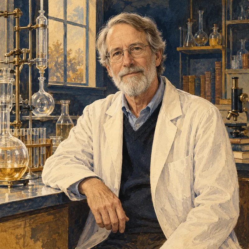
George P. Smith
1941- · United States · Nobel Chemistry 2018
Phage display of peptides and antibodies