The discovery, explained · Chemist no. 210 of 350
Joachim Frank and the Art of Building 3D Molecules from Noisy 2D Pictures
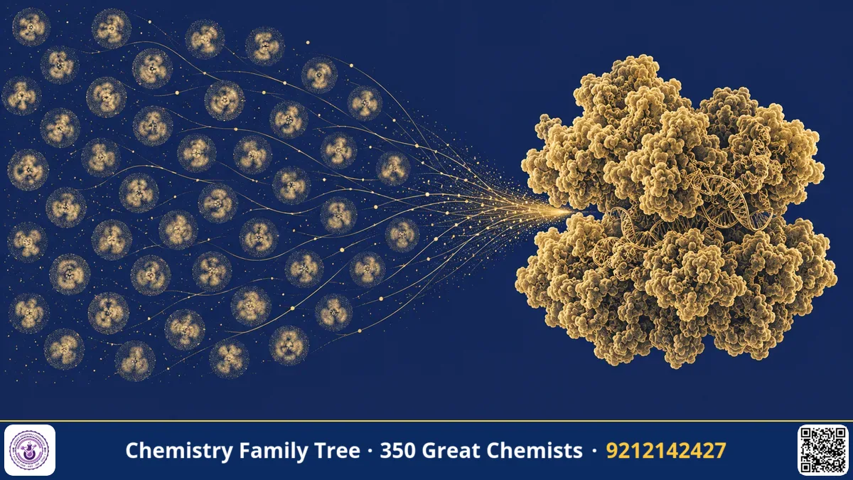
A cryo-electron microscope image of a biomolecule looks like a faint grey smudge. It is extremely noisy, because to avoid destroying the sample, only a tiny dose of electrons can be used. Yet from thousands of such smudges, scientists can compute a sharp three-dimensional model that shows individual atoms. The key computational ideas behind this were developed by Joachim Frank, a German-born biophysicist working in the United States. For this he shared the 2017 Nobel Prize in Chemistry with Jacques Dubochet and Richard Henderson.
The Scientific Problem Before the Discovery
An electron micrograph is a two-dimensional projection of a three-dimensional object, like a shadow. To get the 3D shape, you need views from many angles. For regular crystals, this was solved by diffraction methods. But many important molecular machines, such as the ribosome, do not form good crystals. In solution, they sit in all possible orientations, and each particle is imaged only once, from whatever angle it happens to have. Moreover, the signal is weak compared to noise. A single image cannot be interpreted.
The problem was therefore: how can one combine many noisy images of identical particles, with unknown orientations, into a reliable 3D structure? In the 1970s, computers were slow and image-processing tools were primitive. A new mathematical framework was needed.
The Discovery: Step by Step
Frank started working on this in the 1970s, when he was at the Max Planck Institute in Munich, and continued in Albany, New York, at the Wadsworth Center. The core of his single-particle reconstruction approach has several stages.
- Image collection. Thousands of images of vitrified particles are recorded at low electron dose.
- Alignment. Images of the same type of particle, in the same view, are shifted and rotated so they overlap. Frank introduced correlation-based alignment methods for this.
- Classification and averaging. Using statistical methods such as correspondence analysis (a multivariate statistics technique), Frank's group sorted images into classes of similar views. Averaging the images in a class increases the signal-to-noise ratio. If N images are averaged, the noise falls roughly as 1/√N.
- Finding orientations. The relative angles of different class averages are determined using the "common lines" principle: any two 2D projections of the same 3D object share a common line in Fourier space (the projection-slice theorem).
- 3D reconstruction. Projections with known angles are combined by back-projection into a three-dimensional density map. The map is refined iteratively by comparing it with the raw images.
Frank's group also developed the SPIDER software package, which made these steps available to the community. In the 1990s and after, the group's reconstructions of the Escherichia coli ribosome (initially at low resolution, then improved) were milestones. They showed the ribosome's shape, tunnels, and the positions of transfer RNA, and later captured the ribosome in different functional states, such as the movement of tRNA and ratcheting between the two subunits during translation.
The Chemistry Behind It
- Fourier transform and projection-slice theorem: The Fourier transform of a 2D projection is a central slice of the 3D Fourier transform of the object. This links real-space images to reciprocal space.
- Signal-to-noise ratio: Averaging N independent noisy measurements improves SNR by a factor of √N.
- Resolution: Measured by Fourier shell correlation, which compares two independent half-maps.
- Conformational heterogeneity: Molecules in solution can occupy several states. Classification can sort them, giving "snapshots" of a working machine.
- Ribosome chemistry: Peptide bond formation is catalysed by the ribosomal RNA, and mRNA is read in triplets (codons) by tRNA anticodons.
Many noisy 2D projections → align + classify + average → assign angles (common lines) → back-projection → 3D density map → atomic model
Applications Today
- Antibiotic design: Cryo-EM structures show how antibiotics bind bacterial ribosomes, guiding drug design against resistant bacteria.
- Disease mechanisms: Structures of protein aggregates in neurodegenerative diseases.
- Vaccines: Shape of viral surface proteins such as the coronavirus spike.
- Machine learning: Modern software uses Bayesian statistics and deep learning to refine maps, building on the same logic of alignment and classification.
- Drug targets: Membrane proteins such as ion channels and receptors that resist crystallisation can now be studied.
Exam Corner
- Who shared the 2017 Chemistry Nobel? Dubochet, Frank, Henderson.
- What is the projection-slice theorem? The 2D Fourier transform of a projection equals a central slice of the 3D Fourier transform of the object.
- How does averaging N images affect noise? SNR improves as √N.
- Which biological machine did Frank famously reconstruct? The ribosome.
- Why is single-particle cryo-EM useful? It works for non-crystalline, solution-state molecules.
Common Misconceptions
- "Cryo-EM produces the 3D picture directly." It is computed from many 2D images.
- "Averaging removes all useful differences." Classification keeps different states apart before averaging.
- "Frank invented the microscope." He developed the computational methods; microscope and sample preparation advances came from others.
Quick Revision Box
- Frank developed single-particle reconstruction methods from the 1970s onward.
- Steps: alignment, classification, averaging, angle assignment, back-projection.
- Uses the projection-slice (common line) theorem.
- SPIDER was his image-processing software system.
- Famous for 3D structure of the ribosome in different states.
- Nobel Prize in Chemistry 2017 (shared).
FAQ
Why do we need thousands of particle images?
Each image is very noisy, and many orientations are needed to cover the 3D structure. Averaging improves the signal.
What is back-projection?
A method of reconstructing a 3D object by smearing each 2D projection back across the volume along the direction in which it was recorded and summing them.
Is Frank a chemist?
He is a biophysicist, but the Nobel Prize recognised his contribution to determining molecular structure, which belongs to chemistry.
How can I use this for exam preparation?
Remember cryo-EM as a structure method that does not need crystals, and compare it with X-ray crystallography and NMR.
Published by ABC Chemistry — IIT-JAM | CSIR-NET | GATE Chemistry Coaching. Visit abcchemistry.com
About the chemist
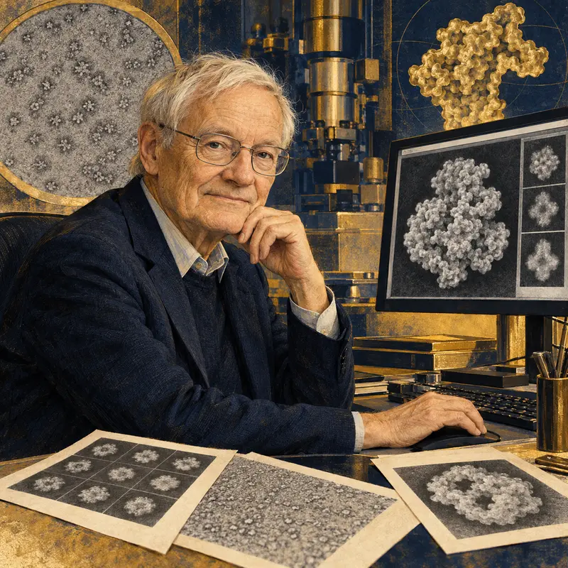
Joachim Frank
1940- · United States · Nobel Chemistry 2017
Image processing for cryo-EM