The discovery, explained · Chemist no. 157 of 350
Johann Deisenhofer and the Photosynthetic Reaction Centre: The First Membrane Protein Structure
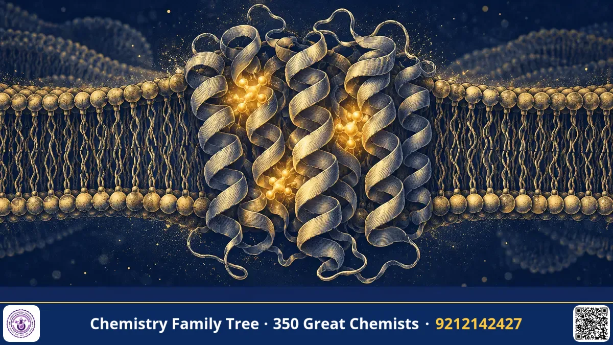
Photosynthesis feeds almost every living thing on Earth, yet until the 1980s nobody had seen the machine that performs its first step: converting a photon of light into separated electric charge. Johann Deisenhofer, together with Robert Huber and Hartmut Michel, determined the three-dimensional structure of the photosynthetic reaction centre of the purple bacterium Rhodopseudomonas viridis by X-ray crystallography. It was the first atomic-level structure of a membrane-bound protein complex. The trio received the 1988 Nobel Prize in Chemistry. For students, this discovery connects protein chemistry, coordination chemistry of porphyrins, electron transfer and X-ray diffraction.
The Scientific Problem Before the Discovery
By the 1970s many soluble proteins, such as myoglobin, lysozyme and haemoglobin, had been solved by X-ray crystallography. Membrane proteins were a different matter. They have hydrophobic surfaces that sit inside the lipid bilayer, and they clump or denature when removed into water. Crystals need identical, regularly packed molecules, and a membrane protein surrounded by a messy shell of lipid does not pack well. As a result, no membrane protein structure was known, and many scientists believed that crystals of these proteins could not be obtained.
At the same time, biochemists knew a great deal about what photosynthetic reaction centres do. Light energy is absorbed by antenna pigments and transferred to a special pair of chlorophyll-type molecules. This pair gives up an electron, which then moves along a chain of cofactors across the membrane. The step creates a charge separation that is later used to make ATP. But how the pigments are arranged in space, and why the electron goes where it does, remained unknown.
The Discovery: Crystals, Diffraction and a Model
The breakthrough began with Hartmut Michel, who, working with Dieter Oesterhelt in Martinsried near Munich, found a way to crystallise the reaction centre from Rh. viridis in 1982. The key was to use a mild detergent that covers the hydrophobic surface of the protein, keeping it soluble while still allowing regular packing, and to add small amphiphilic molecules that helped crystals grow. These were the first well-ordered three-dimensional crystals of a membrane protein.
Deisenhofer then joined Michel and Huber at the Max Planck Institute of Biochemistry in Martinsried to solve the structure. The main steps were:
- Collect diffraction data. The crystals diffracted X-rays well enough to give data to about 3 Å resolution.
- Solve the phase problem. Heavy-atom derivatives of the crystals were prepared, and phases were obtained by multiple isomorphous replacement, a standard method in protein crystallography.
- Compute electron density maps. From the amplitudes and phases, a Fourier transform gave maps of electron density.
- Build the model. Using computer graphics, the team fitted the amino acid chains and pigments into the density, and refined the model against the data.
The results, published in 1984 and 1985, showed a complex of four protein subunits named L, M, H and a cytochrome. The L and M subunits each cross the membrane several times as α-helices (five in each), and they are arranged in a nearly two-fold symmetrical way. The H subunit sits on the cytoplasmic side, and the cytochrome with four haem groups is on the other side. Inside the L and M subunits the team located the cofactors:
- A special pair of bacteriochlorophyll molecules at the periplasmic side.
- Two accessory bacteriochlorophylls.
- Two bacteriopheophytins.
- Two quinones (QA and QB) and a non-haem iron atom between them.
The Chemistry Behind It
The pigments form two similar branches, but electron transfer follows mainly one of them. After the special pair is excited by light (P → P*), it donates an electron:
P* → P⁺ + e⁻ (to the bacteriopheophytin, through the accessory bacteriochlorophyll)
The electron then moves on to QA and finally to QB. The first charge separation takes only a few picoseconds, while later steps take longer. The oxidised special pair P⁺ is refilled with an electron from the cytochrome haem groups. After QB receives two electrons and two protons, it becomes a quinol:
Q + 2e⁻ + 2H⁺ → QH₂
This is the same type of quinone–quinol redox couple seen in organic chemistry and in respiration. The close packing of the pigments, the distances between them (about 10 Å or less), and the surrounding protein environment control the speed and direction of electron transfer. This fits with electron transfer theory, where rate depends exponentially on distance.
Applications Today
- Membrane protein crystallography: The method used by Michel became the template for crystallising receptors, channels and transporters, which are major drug targets.
- Artificial photosynthesis: Chemists design molecular systems that mimic charge separation to produce fuels from sunlight, a theme of solar energy research including work in Indian institutes.
- Solar cell design: Dye-sensitised and organic solar cells borrow ideas of pigment arrangement and electron transfer chains.
- Understanding of biology: The structure helped to explain how plants and cyanobacteria carry out the same step, in photosystem II.
Exam Corner
- The 1988 Nobel Prize in Chemistry went to Deisenhofer, Huber and Michel for the three-dimensional structure of the photosynthetic reaction centre.
- Organism: the purple bacterium Rhodopseudomonas viridis.
- Cofactors: special pair, bacteriochlorophylls, bacteriopheophytins, quinones QA and QB, non-haem iron.
- Technique: X-ray crystallography with heavy-atom derivatives (multiple isomorphous replacement).
- Transmembrane segments are mostly α-helical, with hydrophobic side chains facing the lipid.
Common Misconceptions
- "The reaction centre makes sugar." It does the light-driven charge separation; sugar formation happens later in the Calvin cycle (in plants).
- "Membrane proteins are just like soluble proteins." They have a hydrophobic outer surface, which makes them hard to crystallise.
- "The structure was solved by Michel alone." Michel produced the crystals; Deisenhofer carried out the structure solution and refinement with Huber's group, and all three shared the credit.
Quick Revision Box
- First 3D structure of a membrane protein complex: bacterial reaction centre.
- Crystals obtained by Hartmut Michel in 1982 using detergents.
- Subunits: L, M, H and cytochrome with four haems.
- Electron path: special pair → bacteriopheophytin → QA → QB.
- QB + 2e⁻ + 2H⁺ → QH₂.
- Nobel Prize in Chemistry, 1988, shared by three scientists.
FAQ
Why was this structure such a landmark?
It proved that membrane protein crystals can be grown and solved, and it opened the way to studying receptors, channels and pumps at atomic resolution.
What is the special pair?
It is a pair of closely stacked bacteriochlorophyll molecules that give up an electron after absorbing light energy.
Did Deisenhofer work alone?
No. He worked in the group of Robert Huber, with Hartmut Michel's crystals as the starting point.
Is the Nobel for Chemistry or Medicine?
Chemistry, in 1988.
Published by ABC Chemistry — IIT-JAM | CSIR-NET | GATE Chemistry Coaching. Visit abcchemistry.com
About the chemist
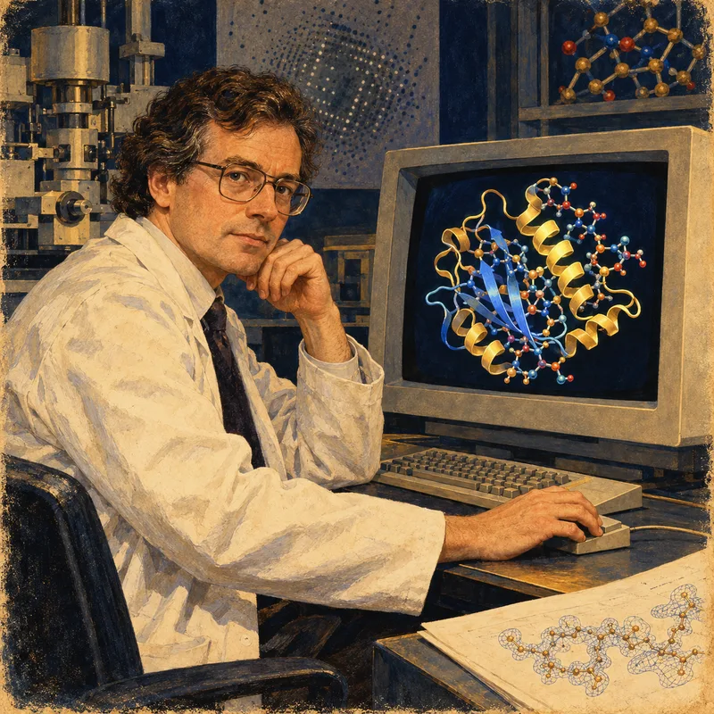
Johann Deisenhofer
1943- · Germany · Nobel Chemistry 1988
Structure of photosynthetic reaction centre