The discovery, explained · Chemist no. 168 of 350
John E. Walker and the Structure of ATP Synthase
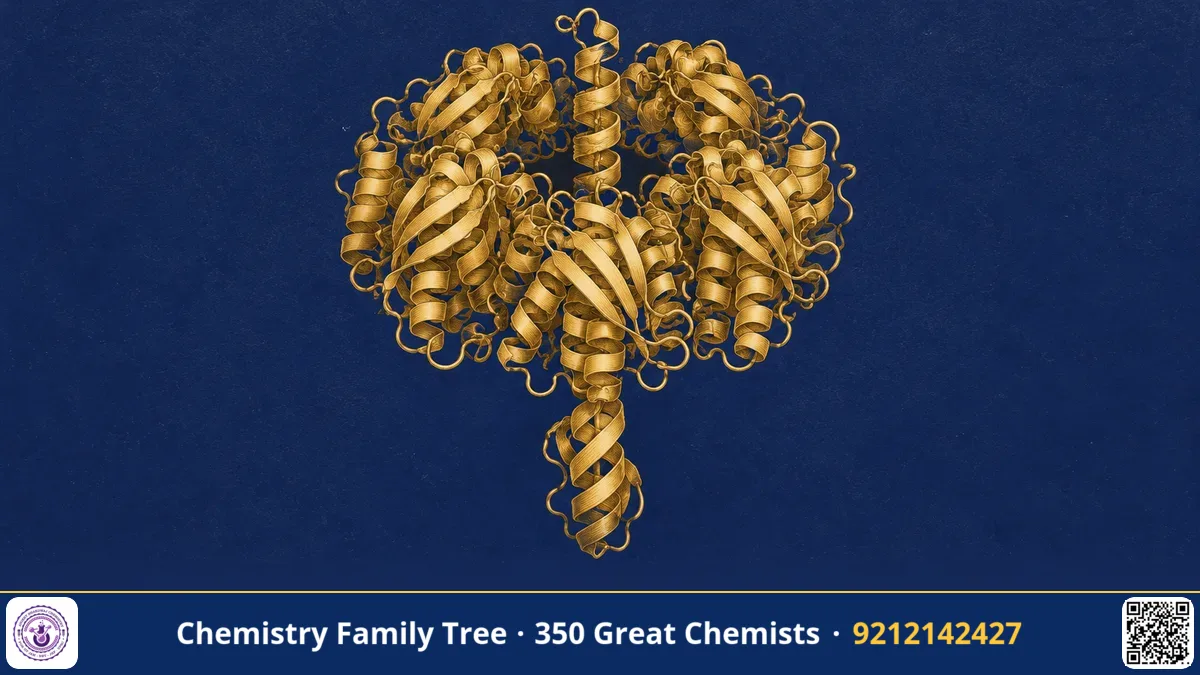
When Paul Boyer proposed his binding change mechanism, it was a bold prediction without a picture. In 1994, the British biochemist John E. Walker and his team at the MRC Laboratory of Molecular Biology in Cambridge provided the picture. They determined the three-dimensional structure of the catalytic head of ATP synthase, called F1-ATPase, at atomic resolution using X-ray crystallography. The structure showed three catalytic subunits in three different states around an asymmetric central shaft. This matched Boyer's mechanism and made rotational catalysis a reality. Walker shared the 1997 Nobel Prize in Chemistry with Boyer; the other half went to Jens Skou. This post explains what Walker did, why it was so difficult and what students must remember.
The Scientific Problem Before the Discovery
By the 1980s, scientists knew that ATP synthase is a large, complex enzyme with a membrane part (Fo) and a soluble part (F1). Biochemists had purified it, but its structure was unknown. The main reaction, shown in simple form, is:
ADP + Pi + n H⁺(outside) ⇌ ATP + H₂O + n H⁺(inside)
Boyer's binding change mechanism said that three catalytic sites alternate between open, loose and tight states, driven by rotation. But there was no direct evidence of the three different states. Also, the amino acid sequences of the many subunits were not known. Without the sequences, a crystallographer cannot build a model into electron density. Two big problems therefore had to be solved: determine the sequences of the proteins, then crystallise a very large and flexible complex.
The Discovery: Step by Step
Walker's research career in the 1970s and 1980s was built around protein chemistry and sequencing. His work had several stages:
- Sequencing of subunits. Walker and his group worked out the amino acid sequences of the subunits of ATP synthase from bovine heart mitochondria, and also from Escherichia coli. In 1982 his group reported that subunits of ATP synthase contain a common nucleotide-binding sequence motif. The sequence motifs are now known as Walker A and Walker B motifs.
- Purification and crystallisation. The F1 part from bovine heart mitochondria was purified in large amounts. After many attempts, crystals suitable for diffraction were obtained, with Walker's colleague Jan Pieter Abrahams and others.
- X-ray diffraction. The crystals were exposed to X-rays and the diffraction pattern was recorded. By solving the phase problem, an electron density map was calculated, and the model of the protein was built into the map.
- Structure published. In 1994 the structure was reported in Nature at 2.8 Å resolution by Abrahams, Leslie, Lutter and Walker.
What the structure showed
The F1 head has the shape of an orange cut into segments. Three α subunits and three β subunits alternate around a central cavity. A long coiled coil formed by the γ subunit passes through the centre. Because the γ subunit is not symmetric, the three β subunits are in different environments. In the crystal, they were found in three states:
- βTP: bound with a non-hydrolysable ATP analogue (AMP-PNP), the tight state
- βDP: bound with ADP, the loose state
- βE: empty, the open state
This was the direct structural evidence for the three states in Boyer's mechanism. The picture also suggested that rotation of γ would cause each β subunit to pass through each state in turn. Later, in 1997, single-molecule experiments by Noji and co-workers directly showed this rotation.
The Chemistry Behind It
Nucleotide binding: the Walker motifs
The Walker A motif (also called the P-loop) has the consensus sequence GXXXXGK[T/S]. It wraps around the phosphate groups of ATP or GTP and coordinates Mg2+. The Walker B motif is a hydrophobic strand ending in an aspartate, which helps to coordinate the Mg2+ ion and the water molecule that attacks the terminal phosphate. These motifs are found in a huge number of ATP- and GTP-binding proteins, such as G proteins, helicases and many ATPases, so they are very important for biochemistry exams.
The hydrolysis step
In the enzyme working in reverse (ATP hydrolysis), a water molecule attacks the γ-phosphorus of ATP in an in-line SN2-type reaction, and the P–O bond to ADP breaks:
ATP + H₂O → ADP + Pi ΔG°′ ≈ −30.5 kJ/mol
Inside the cell, the actual free energy change is more negative because the concentrations of ATP, ADP and Pi are far from standard.
The whole machine
Walker's group later also studied the peripheral stalk and the c-ring of the Fo part. The number of c-subunits varies among species, and it decides how many protons are used per ATP. For example, the bovine mitochondrial enzyme has eight c-subunits, so eight protons are needed for each full rotation that makes three ATP, about 2.7 H+ per ATP. The ratio is different in bacteria or chloroplasts.
Applications Today
- Drug design: Knowing the structure helps to develop inhibitors. Bedaquiline, used against drug-resistant tuberculosis, targets the c-ring of the mycobacterial ATP synthase.
- Understanding mitochondrial disease: Mapping mutations on the structure explains how they disturb ATP synthesis.
- Structural biology method: The work is a landmark case of X-ray crystallography for a large membrane-associated assembly. Later work used cryo-electron microscopy.
- Bioinformatics: The Walker A and B motifs are used to predict ATP- and GTP-binding proteins from genome sequences.
- Synthetic biology: Rotary motors based on ATP synthase inspire the design of nanomachines.
Exam Corner
- Walker A motif: GXXXXGK[T/S], the P-loop that binds phosphates of ATP. Walker B motif: ends with an acidic residue (Asp), binds Mg2+.
- The 1994 F1 structure showed three β subunits in different nucleotide states, confirming Boyer's binding change mechanism.
- The γ subunit is asymmetric; it is the rotor that causes conformational change.
- F1 composition is α3β3γδε. Catalytic sites are mainly on β subunits.
- Nobel Prize in Chemistry 1997 was shared by Walker, Boyer and Skou (Boyer and Walker for ATP synthesis; Skou for the Na+,K+-ATPase).
Common Misconceptions
- "Walker discovered ATP synthase." The enzyme was known well before him. Walker determined its sequence and structure.
- "The catalytic sites are in the membrane." The catalytic sites are in the F1 head outside the membrane. The membrane part, Fo, carries protons.
- "Walker and Boyer did the same experiment." Boyer worked from kinetics and isotope exchange to predict a mechanism. Walker used structure to show it physically.
Quick Revision Box
- 1994: X-ray structure of bovine F1-ATPase at 2.8 Å.
- Three β subunits in three states: with AMP-PNP, with ADP, and empty.
- Asymmetric γ coiled coil is the central rotor.
- Walker A (P-loop) and Walker B motifs bind nucleotides and Mg2+.
- Rotation proven directly in 1997 by Noji, Yoshida, Kinosita and colleagues.
- Nobel Prize in Chemistry 1997 with Boyer and Skou.
FAQ
What is the Walker A motif?
It is a short sequence in many nucleotide-binding proteins that forms a loop to hold the phosphate groups of ATP or GTP. Its consensus is GXXXXGK[T/S].
Why was the structure so important?
It gave physical proof for three catalytic states and the asymmetry of the central shaft, turning a prediction into an observation.
How is Walker's work linked to Boyer's?
Boyer predicted that three sites cycle through different states with rotation. Walker's structure showed three different sites and an asymmetric shaft in a single crystal.
Which technique did Walker use?
X-ray crystallography, supported by protein sequencing and biochemical purification of the enzyme from bovine heart mitochondria.
Published by ABC Chemistry — IIT-JAM | CSIR-NET | GATE Chemistry Coaching. Visit abcchemistry.com

About the chemist
John E. Walker
1941- · United Kingdom · Nobel Chemistry 1997
Structure of ATP synthase