The discovery, explained · Chemist no. 164 of 350
Michael Smith and Site-Directed Mutagenesis: Editing a Protein One Amino Acid at a Time
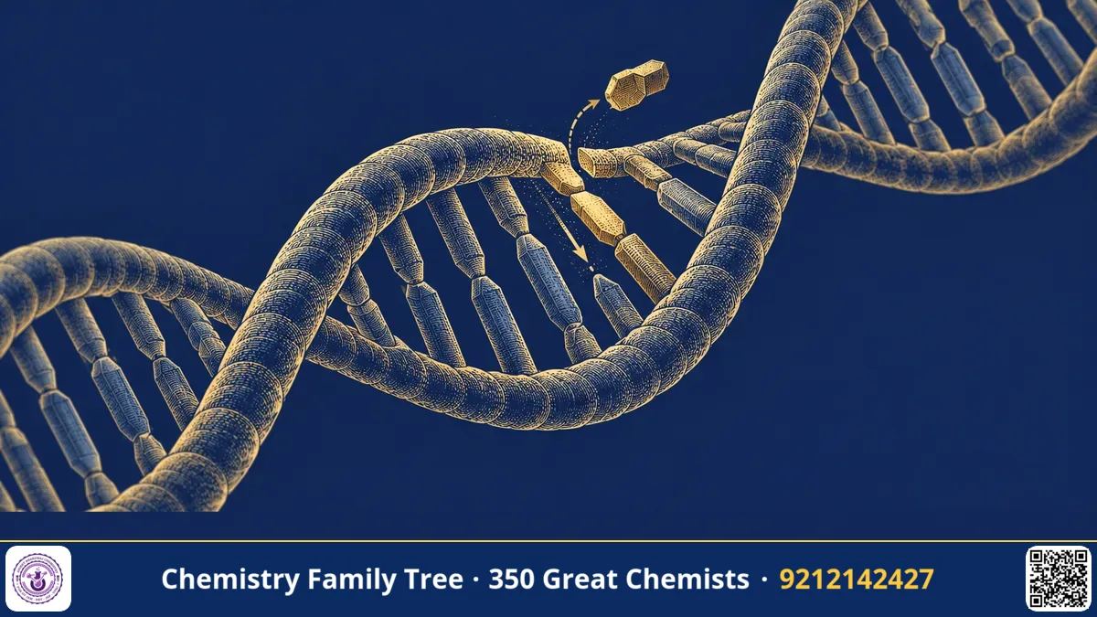
Suppose you want to know what a single amino acid does inside an enzyme. Which residue grips the substrate? Which one acts as an acid or a base? One way to find out is to change that residue to something else and see what happens. Before 1978, researchers could only wait for random mutations or create them with chemicals and radiation, then search through thousands of mutants. Michael Smith developed a method to introduce a chosen change at a chosen position in a gene. It is called oligonucleotide-directed site-directed mutagenesis. It gave chemists and biologists a molecular "find and replace" for proteins. For this contribution Smith shared the 1993 Nobel Prize in Chemistry with Kary Mullis.
The Scientific Problem Before the Discovery
The genetic code links a DNA sequence to a protein sequence. Three bases (a codon) code for an amino acid; for example, the codon GAA codes for glutamic acid, and a change to GAU would give aspartic acid. In principle, changing one base changes one amino acid. In practice, however, researchers had no way to make a specific change. Mutagens such as ultraviolet light, X-rays and chemicals like nitrous acid cause random changes. To study a particular residue, you would have to screen enormous numbers of organisms.
At the same time, chemists could now synthesise short DNA strands of defined sequence (oligonucleotides), thanks to the work of Har Gobind Khorana and others. Smith, who had worked with Khorana, realised that a synthetic oligonucleotide could be used as a precise tool to direct a mutation.
The Discovery: A Mismatched Primer
In the late 1970s, working at the University of British Columbia in Vancouver, Smith and his colleagues, including Clyde Hutchison and Marshall Edgell, used the small single-stranded DNA of bacteriophage φX174 as a test system. They had a mutant phage with a known single-base change, and wanted to show that a synthetic primer could correct it, or create a new change. The strategy was as follows.
- Prepare single-stranded circular DNA. The gene to be changed is carried in a single-stranded circular phage DNA.
- Design a synthetic oligonucleotide. A short DNA (about 15–20 bases) is made which is complementary to the region of the gene, except at one position where it deliberately has a different base. This is a mismatched primer.
- Anneal. The primer binds to the template at the right place, forming a double helix with one mismatched base pair that sticks out of the helix.
- Extend and seal. DNA polymerase extends the primer around the circle, using the template, and DNA ligase joins the ends, giving a fully double-stranded circular molecule. One strand carries the mutation and the other the original sequence.
- Replicate in cells. When the DNA enters bacteria and replicates, each strand directs the formation of its own progeny, and a fraction of the progeny carry the desired mutation.
- Screen and confirm. Mutant plaques or clones are identified (for example by hybridisation with the same oligonucleotide) and the change is verified by sequencing.
The first demonstration was published in 1978 with the φX174 system. Later, the method was improved with cloning vectors such as M13 phage, and with selection tricks that raised the yield of mutants from a few percent to a large majority.
The Chemistry Behind It
The method depends on base-pair stability. A primer with one mismatch in the middle still anneals, because 15 or more correctly paired bases give sufficient stability. A mismatch at the 3′ end would not be extended efficiently, so the mismatch is placed near the centre.
| Type of change | Example |
|---|---|
| Substitution | Codon change from GAA (Glu) to GAU (Asp) or to GCA (Ala) |
| Insertion | Extra codons added to insert a new residue or a tag |
| Deletion | Removal of codons to delete a segment |
A common application is alanine scanning, in which one residue after another is replaced with alanine, to find which side chains are important for function. In enzymology, replacing the nucleophilic serine of a protease, Ser → Ala, destroys its activity, confirming the catalytic role. The kinetic parameters kcat and Km of mutants are compared with the wild type to find out whether binding or catalysis was affected:
v = kcat[E]₀[S] / (Km + [S])
Applications Today
- Enzyme mechanism studies: Testing the role of individual residues in the active site.
- Protein engineering: Designing enzymes that are more stable at high temperature, such as the ones used in detergents and industry.
- Drug development: Making modified therapeutic proteins, such as insulin analogues with faster or slower action, and antibodies with improved binding.
- Disease research: Recreating a patient mutation in the laboratory to study how it causes disease.
- Agriculture: Modifying plant proteins to improve traits, as part of modern biotechnology.
- Research in India: Widely used in biotechnology and structural biology laboratories in IITs, IISc and CSIR institutes.
Exam Corner
- Michael Smith shared the 1993 Nobel Prize in Chemistry with Kary Mullis, for oligonucleotide-based, site-directed mutagenesis.
- A mismatched synthetic oligonucleotide primer introduces a specific base change.
- Steps: anneal primer, extend with polymerase, ligate, transform, screen, sequence.
- Alanine scanning identifies functionally important residues.
- Smith worked with Khorana, the 1968 Nobel laureate in Physiology or Medicine, who pioneered oligonucleotide synthesis.
Common Misconceptions
- "Site-directed mutagenesis uses radiation." It uses a synthetic DNA primer to make a precise change, not random damage.
- "It is the same as PCR." Both use primers, but PCR amplifies DNA; mutagenesis changes the sequence. Modern protocols often combine the two.
- "Every mutation is harmful." Many changes are neutral, and some improve a property; the point is to test hypotheses.
Quick Revision Box
- Method: mismatched oligonucleotide primer directs a specific change.
- First shown in 1978 with bacteriophage φX174 DNA.
- Steps: anneal, extend, ligate, transform, screen, sequence.
- Uses: enzyme mechanism, protein engineering, therapeutic proteins.
- Alanine scanning replaces residues with Ala.
- 1993 Nobel Prize in Chemistry with Kary Mullis.
FAQ
Why is a mismatch placed in the middle of the primer?
So that enough correct base pairs flank it on both sides to hold the primer in place, and the 3′ end remains correctly paired for extension.
How did Smith relate to Khorana?
Smith worked as a postdoctoral researcher with Har Gobind Khorana in Vancouver, where he learned oligonucleotide chemistry.
Is the method still used?
Yes, in improved forms. It remains a standard tool, along with newer genome-editing methods.
Was his Nobel in Chemistry or Medicine?
Chemistry, 1993.
Published by ABC Chemistry — IIT-JAM | CSIR-NET | GATE Chemistry Coaching. Visit abcchemistry.com

About the chemist
Michael Smith
1932-2000 · Canada · Nobel Chemistry 1993
Site-directed mutagenesis