The discovery, explained · Chemist no. 211 of 350
Richard Henderson and the Structure of Bacteriorhodopsin
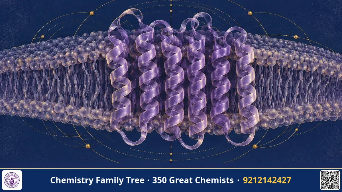
Membrane proteins sit in the fatty membranes of cells and control what goes in and out. They are the targets of more than half of all modern drugs, yet they were extremely difficult to study, because they refuse to crystallise in water. In 1975, Richard Henderson and Nigel Unwin used an electron microscope to obtain the first three-dimensional picture of a membrane protein, bacteriorhodopsin, at a resolution that showed its helices. By 1990 Henderson and colleagues had a near-atomic model. This work proved that electron microscopy could reveal biological structure at high resolution. Henderson shared the 2017 Nobel Prize in Chemistry with Jacques Dubochet and Joachim Frank.
The Scientific Problem Before the Discovery
Before the 1970s, protein structures were determined mostly by X-ray crystallography, which requires well-ordered 3D crystals. Soluble proteins such as myoglobin and lysozyme could be crystallised, but membrane proteins, which need a lipid environment, were stubborn. Taking them out of the membrane with detergents often made them unstable.
Electron microscopy had a different problem: damage. Electrons are much more damaging to biological material than X-rays per unit of information, and the high vacuum dries the specimen. To get any detail, one needed a specimen that was very regular, so that thousands of identical molecules could be averaged, and a method to protect it in the microscope.
The Discovery: Step by Step
- Find a natural 2D crystal. The halophilic archaeon Halobacterium salinarum (formerly H. halobium) makes a protein, bacteriorhodopsin, which is packed in a hexagonal lattice in patches of membrane called the purple membrane. These patches are natural two-dimensional crystals.
- Protect the specimen. Henderson and Unwin embedded the purple membrane in glucose, which replaces water and keeps the structure intact in the vacuum. They used a very low electron dose to minimise damage.
- Record electron diffraction and images. Electron diffraction gave very accurate amplitudes of structure factors, and images gave phases. Combining amplitudes from diffraction with phases from images in Fourier space is a key trick.
- Tilt the specimen. Images at different tilt angles provided views from different directions, enabling a 3D reconstruction by Fourier methods.
- Interpret the map. In 1975 the 7 angstrom map showed seven rod-like transmembrane alpha helices, a bundle running across the membrane. The later 1990 model resolved the protein to near-atomic detail (about 3.5 angstrom in-plane) and allowed an atomic model of the polypeptide chain.
This was the first time the structure of a membrane protein was seen, and it also provided the first evidence for the seven-helix architecture that is shared by a huge family of receptors, including G-protein-coupled receptors (GPCRs).
The Chemistry Behind It
- Retinal and the Schiff base: Bacteriorhodopsin binds the chromophore all-trans-retinal through a protonated Schiff base (an imine) to the side chain of Lys216. The chromophore gives the purple colour, with absorption near 570 nm.
- Photocycle: Absorption of a photon isomerises retinal from all-trans to 13-cis. The Schiff base then passes its proton to a nearby aspartate (Asp85), and the protein goes through intermediates (K, L, M, N, O) before returning to the starting state.
- Proton pumping: Each cycle moves a proton from the inside to the outside of the cell, building a proton gradient that drives ATP synthesis. This is light energy converted to chemical potential energy.
- Helix packing: Seven hydrophobic transmembrane helices cross the lipid bilayer (about 30 angstrom thick), a typical pattern for integral membrane proteins.
- Fourier methods: A structure is the Fourier transform of diffraction data with phases. Phases are the hard part, and electron microscopy gives them directly from images.
retinal (all-trans) + hv → 13-cis retinal → H⁺ transfer to Asp85 → H⁺ released outside → reprotonation from inside → all-trans
Applications Today
- Drug discovery: Understanding GPCR-like seven-helix proteins guides drug design.
- Optogenetics: Related microbial rhodopsins, such as channelrhodopsin, are used to control nerve cells with light.
- Bioenergetics teaching: Bacteriorhodopsin is a classic model for proton pumps and chemiosmosis.
- Cryo-EM: Henderson later championed direct electron detectors that made single-particle cryo-EM practical for small proteins.
- Bio-inspired devices: Research on light-driven biomolecular energy converters.
Exam Corner
- What is the purple membrane? Natural 2D crystalline patches of bacteriorhodopsin in Halobacterium.
- Chromophore and linkage: all-trans retinal bound as a protonated Schiff base to a lysine.
- Function of bacteriorhodopsin: light-driven outward proton pump.
- How many transmembrane helices? Seven.
- 2017 Nobel in Chemistry: Henderson, Dubochet, Frank.
Common Misconceptions
- "Bacteriorhodopsin is related to chlorophyll." It is not; it uses retinal and has no photosynthetic electron transport chain.
- "It is found in human eyes." Human vision uses rhodopsin, which is a similar seven-helix protein with retinal but a different function.
- "Electron microscopy was always low resolution." This work showed it could approach atomic detail.
Quick Revision Box
- Henderson and Unwin published the first membrane protein structure in 1975.
- Sample: purple membrane of Halobacterium, glucose-embedded.
- Seven transmembrane alpha helices; retinal Schiff base chromophore.
- Light-driven proton pump: all-trans to 13-cis isomerisation.
- Near-atomic electron crystallography model in 1990.
- Nobel Prize in Chemistry 2017 (shared).
FAQ
What is a Schiff base?
An imine formed by condensation of an amine (here, lysine) with an aldehyde (retinal): R-CH=N-R'.
Why did the purple membrane help?
Its natural 2D crystal order allowed averaging over thousands of molecules, overcoming the weak signal.
How does it connect to GPCRs?
Both have seven transmembrane helices, although they are not evolutionarily related in sequence; the shared fold became a model for many receptors.
Is this topic relevant for exams?
Yes, for bioinorganic and biochemistry-related questions on proton pumps, retinal photochemistry and structure determination methods.
Published by ABC Chemistry — IIT-JAM | CSIR-NET | GATE Chemistry Coaching. Visit abcchemistry.com
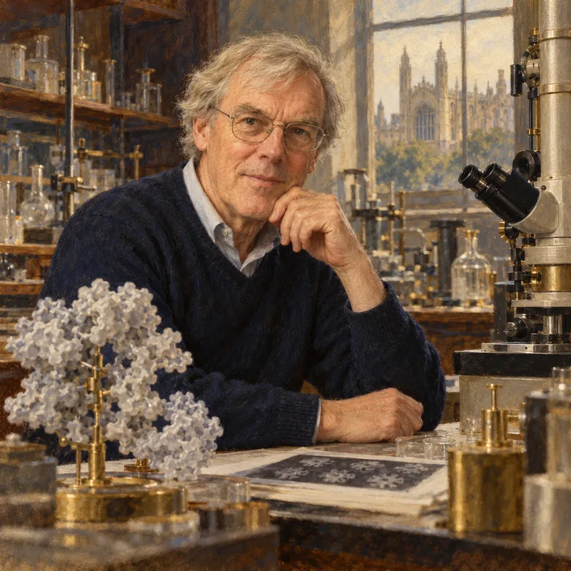
About the chemist
Richard Henderson
1945- · United Kingdom · Nobel Chemistry 2017
Cryo-EM of biomolecules